Foot fractures and dislocations
Peer reviewed by Dr Colin Tidy, MRCGPLast updated by Dr Laurence KnottLast updated 17 Sept 2021
Meets Patient’s editorial guidelines
Medical Professionals
Professional Reference articles are designed for health professionals to use. They are written by UK doctors and based on research evidence, UK and European Guidelines. You may find the Broken toe article more useful, or one of our other health articles.
In this article:
See also the separate articles Painful Foot and Heel Pain.
Approximately 10% of all fractures occur in the bones of the foot. These bones include:
Hindfoot: the calcaneus and the talus.
Midfoot: the navicular, the cuboid and 3 cuneiforms.
Forefoot: 5 metatarsals and 14 phalanges.
The foot also contains sesamoid bones (bones embedded within a tendon).
Severe injuries to the foot can result in significant long-term pain and loss of function. Multiple fractures or dislocations of the feet are often initially overlooked in cases of multiple, severe trauma. Outcomes are worse if treatment is not immediately initiated, if patients subsequently had neuritis or reflex sympathetic dystrophy, or if patients were involved in ongoing related litigation1 . It has been thought that delayed soft-tissue coverage was associated with an adverse prognosis but this was not substantiated in one study2 .
Stress fractures are common in athletes, and may occur in every bone of the foot and ankle, except the smaller toes3 .
Initial management includes ice, immobilisation and elevation. Any delay in providing adequate specific treatment increases the risk of post-traumatic osteoarthritis. Other potential complications include non-union, avascular necrosis, compartment syndromes, vascular injuries, post-traumatic ankle deformities and tarsal tunnel syndrome.
Continue reading below
Investigations
X-rays
The Ottawa foot rules help to predict significant midfoot fractures. A BMJ review of sensitivity of the rules as a predictor suggested that the tool has an almost 100% sensitivity in adults and children.
The Ottawa rules suggest that X-rays are required if any of the following are present4 5 :
Point tenderness over the base of the fifth metatarsal.
Point tenderness over the navicular bone.
Inability to take four steps, both immediately after injury and when seen for assessment.
A separate review in 2003 suggested that the rules may have a lower sensitivity (93%) when applied to children, but with a reassuringly high negative predictive value of 95%6 .
Scans
Bone scans, CT scans, MRI and ultrasound may help to diagnose certain foot fractures that are not seen on plain X-rays.
Talar injuries7 8
Falls on to the feet or violent dorsiflexion of the ankle (eg, against car pedals in a car accident) may cause fractures to the anterior body or articular dome of the talus.
Talar fracture is the second most common fracture of the tarsal bones.
Talar injuries are not easily diagnosed and can create significant long-term disability when missed. CT scanning is extremely helpful in diagnosing and treating them.
Displaced fractures require open reduction and internal fixation.
Prolonged non-weight-bearing and immobilisation are needed.
Avascular necrosis and post-traumatic arthritis to the subtalar and tibiotalar joints are unfortunately frequent complications.
Neck and body fracture
This is the most common type of talar fracture:
It may be associated with subtalar dislocation.
Non-displaced fractures are treated with a non-weight-bearing short leg cast.
Displaced fractures usually require surgical fixation.
Lateral process fracture
Increasingly common because of snowboarding injuries.
Treatment includes immobilisation with avoidance of weight-bearing.
Posterior process (Shepherd's) fracture
Caused by damage to the posterior process of the talus, usually as a result of sudden plantar flexion or repetitive motion, especially dancing or kicking.
Clinical examination is usually nonspecific and plain X-rays normal.
Treatment includes immobilisation with either partial or full weight-bearing.
Transchondral/osteochondral talar dome fracture
Rare; often presents as a non-healing ankle sprain. There is tenderness of the talar dome with the foot in dorsiflexion.
May be clinically indistinguishable from an ankle sprain and plain X-rays may be normal. A bone scan may be required.
Delayed presentation may include crepitus, joint locking and laxity of lateral and anterior ankle ligaments.
Initial management involves immobilisation without weight-bearing.
Dislocation of the talus9
Rare; usually results from very high-energy trauma.
Peritalar and subtalar dislocations involve the articulation between the talus and calcaneum. Midtarsal dislocations involve the midtarsal joint (between the calcaneum and talus posteriorly and the navicular and cuboid anteriorly).
The dislocation is often open and results in avascular necrosis of the talus and arthritis; post-injury joint infection is the single most important factor leading to poor outcomes.
Open reduction and internal fixation are required.
Continue reading below
Calcaneal fractures10
Most calcaneal fractures follow a fall from height directly on to the heels. Calcaneal fractures are often bilateral.
Optimal management of calcaneal fractures is controversial, as correlation between anatomical restoration and outcome is not proven, and complications after surgery are frequent.
Falls from a height usually result in multiple associated injuries - eg, lumbar compression fractures, forearm fractures, and ankle, femur and elbow fractures. There should also be a high index of suspicion for thoracic aortic rupture and renal vascular pedicle disruption.
Calcaneal fractures are divided into intra-articular and extra-articular fractures on the basis of subtalar joint involvement11 :
Intra-articular joint depression fractures
These are the most common type of calcaneal fracture:
Lateral foot X-rays show breaks in the cortices, trabeculae or signs of compression (reduction in Böhler's angle)12 13 . Böhler's angle is the posterior angle formed by intersection of a line from the posterior to the middle facet and a line from the anterior to the middle facet; Böhler's angle is normally between 20° and 40°. Angles less than 20°, or more than 5° smaller than that of the uninjured side, indicate a fracture.
Open reduction and internal fixation is usually necessary. However, one study suggests that minimally invasive surgery reduces the risk of serious wound complications14 .
Extra-articular calcaneal fractures
Extra-articular fractures account for 30% of all calcaneal fractures in adults.
Initial management includes a compression dressing, rest, ice and elevation, with orthopaedic follow-up.
Navicular injuries
Navicular fractures are rare. They are most often stress fractures, occurring in young athletes15 .
They usually heal well with immobilisation and weight-bearing as tolerated.
Displaced fractures involving the navicular body have a high incidence of avascular necrosis and require open reduction and internal fixation.
Complete dislocation of the navicular is rare and prompt reduction under general anaesthetic is required. It may require open reduction and arthrodesis16 .
Continue reading below
Fractures at the Lisfranc (tarsometatarsal) joint
The Lisfranc joint is the area of articulation between the midfoot and forefoot: it is therefore composed of the five tarsometatarsal joints. Foot fractures and traumatic ligament injuries can result in deformity, instability, pain and degenerative disease of the Lisfranc joint17 .
Although injuries to the Lisfranc ligament complex have been associated with high-energy trauma (eg, motor vehicle collisions), they can also result from low-energy trauma, including leisure activities or athletic activity18 .
Tarsometatarsal dislocation can be easily missed on standard foot X-rays. It can result in post-traumatic arthritis and reflex sympathetic dystrophy. To facilitate diagnosis, grasp the first and second metatarsals and move them alternately through plantar flexion and dorsiflexion.
CT imaging is useful if clinical suspicion is high, as plain X-rays can appear normal.
Fractures at the Lisfranc joint generally require urgent open reduction and fixation.
Metatarsal fractures19 20
Metatarsal fractures are relatively common.
If malunited they lead to pain and disability.
Multiple metatarsal fractures may be caused by direct trauma (eg, a heavy object falling on to the foot) or crush injuries (eg, a vehicle wheel).
Management includes analgesia, support in a plaster of Paris (POP) backslab, manipulation under anaesthesia, K-wire fixation or, occasionally, open reduction and internal fixation. It is important to check and monitor the dorsalis pedis pulse.
Non-displaced fractures and fractures of the second to fourth metatarsal with displacement in the horizontal plane can be treated conservatively with protected weight-bearing in a cast shoe for 4-6 weeks.
In most displaced fractures internal fixation is needed.
Percutaneous pinning is suitable for most fractures of the metatarsals. Fractures with joint involvement and multiple fragments frequently require open reduction and plate fixation.
The metatarsals are the most common site of stress fractures.
First metatarsal fracture
The least commonly fractured metatarsal.
Minimally displaced or non-displaced fractures: management usually involves immobilisation without weight-bearing. Displaced fractures usually require open reduction and internal fixation.
Second, third and fourth metatarsals
Fractures are very common.
Non-displaced and displaced fractures usually heal well, with weight-bearing as tolerated in a cast, rigid orthopaedic shoe or elastic support bandages.
Disruptions of the Lisfranc joint must be excluded.
Fifth metatarsal fractures21
The proximal fifth metatarsal is the most common site of midfoot fracture. Fractures are generally of two types:
Proximal avulsion fracture
Fractures at the proximal tuberosity are very common and termed pseudo-Jones or tennis fractures (mid-shaft and distal fractures are much less common). They are usually associated with a lateral ankle strain and often follow inversion injuries of the ankle.
This area should always be checked in patients with ankle injuries, and foot X-rays requested if tender.
Accessory bones or the apophysis (runs parallel to the fifth metatarsal base) may cause confusion when interpreting X-rays.
Treatment includes analgesia, elevation and support in a padded crepe bandage, or a below-knee POP if symptoms are severe.
Usually heals well with a compression dressing and weight-bearing as tolerated.
May require open reduction and tension-band wiring or screw fixation if displaced more than 2 mm or with more that 30% of the joint involved.
Jones fracture
Less common; this is a transverse fracture at the metaphyseal-diaphyseal junction of the fifth metatarsal. Treatment involves an individualised approach tailored to the level of activity and time to union.
Displacement tends to increase with continued weight-bearing.
Initial therapy includes analgesia and immobilisation without weight-bearing.
Frequently requires surgical intervention22 .
Prone to non-union, requiring bone grafting and internal fixation.
Metatarsal stress fractures3 23
A stress fracture is a partial or complete fracture caused by repeated application of stress lower than the stress required to fracture the bone in a single loading.
The most common site is the second metatarsal shaft, but the third metatarsal or, more rarely, other metatarsals may be affected.
May present with swelling over the forefoot and localised tenderness over the metatarsal shaft. Longitudinal compression of the metatarsal shaft (pressing on the metatarsal head below the toe) causes pain along the metatarsal shaft.
Otherwise healthy athletes, especially runners, sustain stress injuries or fractures.
They are not considered fragility fractures, although osteoporosis is a predisposing factor.
Stress fractures account for 0.7% to 20% of all sports medicine clinic injuries.
Track-and-field athletes have the highest incidence of stress fractures compared with other athletes: The sites of stress fractures vary from sport to sport (eg, among track athletes, stress fractures of the navicular, tibia and metatarsal are common; in distance runners, the tibia and fibula; in dancers, the metatarsals).
In the military, the calcaneus and metatarsals are the most common sites, especially in new recruits, due to the sudden increase in running and marching without adequate preparation.They are referred to as march fractures.
Presentation is often subtle, so a high index of suspicion is needed.
X-rays are often initially normal (and remain normal in up to half of all cases). An isotope bone scan may be required for diagnosis, although MRI is also used. Callus or periosteal reaction is sometimes seen on X-rays after 2-3 weeks.
Treatment is symptomatic with analgesia, elevation, rest and reduced activity as required. A padded insole may help. Firm shoes or boots tend to be more comfortable. A below-knee POP or Aircast® boot may be required if the patient is unable to bear weight.
Aircast walking boot
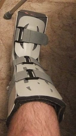
By Pagemaker787, via Wikimedia Commons
Stress fractures of the first to fourth metatarsal shafts typically heal well with rest alone. Full recovery can be expected within 6-8 weeks.
'High-risk' stress fractures include the medial malleolus, the talus, the navicular bone, the base of the fifth metatarsal, and the hallux sesamoids.
Toe fractures 20
Fractures of the toe are among the most common lower limb fractures.
They are most frequently caused by a crush injury or axial force such as stubbing.
Joint hyperextension and stress fractures are less common.
Most patients have point tenderness at the fracture site or pain with gentle axial loading.
X-rays usually identify fractures, determining displacement, and evaluating adjacent phalanges and digits.
Referral is indicated in patients with circulatory compromise, open fractures, significant soft tissue injury, fracture-dislocations, displaced intra-articular fractures, or fractures of the first toe that are unstable or involve more than 25% of the joint surface.
Most children with fractures of the epiphysis should be referred.
Stable, non-displaced toe fractures should be treated with buddy taping (strapping the fractured toe to an adjacent uninjured toe) and a rigid-sole shoe to limit joint movement.
Patients with displaced fractures of the first toe may require referral for reduction and rigid immobilisation. Displaced fractures of the lesser toes should be treated with reduction and buddy taping. Irreducible fractures may require open reduction and internal fixation.
Union occurs in 3-8 weeks but symptoms usually improve much earlier.
Dr Mary Lowth is an author or the original author of this leaflet.
Further reading and references
- Foot Menu; Wheeless' Textbook of Orthopaedics
- Myerson MS, McGarvey WC, Henderson MR, et al; Morbidity after crush injuries to the foot. J Orthop Trauma. 1994 Aug;8(4):343-9.
- Steiert AE, Gohritz A, Schreiber TC, et al; Delayed flap coverage of open extremity fractures after previous vacuum-assisted closure (VAC) therapy - worse or worth? J Plast Reconstr Aesthet Surg. 2009 May;62(5):675-83. doi: 10.1016/j.bjps.2007.09.041. Epub 2008 Mar 25.
- Brockwell J, Yeung Y, Griffith JF; Stress fractures of the foot and ankle. Sports Med Arthrosc. 2009 Sep;17(3):149-59.
- Stiell I; Ottawa ankle rules. Can Fam Physician. 1996 Mar;42:478-80.
- Bachmann LM, Kolb E, Koller MT, et al; Accuracy of Ottawa ankle rules to exclude fractures of the ankle and mid-foot: systematic review. BMJ. 2003 Feb 22;326(7386):417.
- Clark KD, Tanner S; Evaluation of the Ottawa ankle rules in children. Pediatr Emerg Care. 2003 Apr;19(2):73-8.
- Bykov Y; Fractures of the talus. Clin Podiatr Med Surg. 2014 Oct;31(4):509-21. doi: 10.1016/j.cpm.2014.06.004. Epub 2014 Aug 13.
- Early JS; Talus fracture management. Foot Ankle Clin. 2008 Dec;13(4):635-57. doi: 10.1016/j.fcl.2008.08.005.
- Burston JL, Isenegger P, Zellweger R; Open total talus dislocation: clinical and functional outcomes: a case series. J Trauma. 2010 Jun;68(6):1453-8. doi: 10.1097/TA.0b013e3181d03b73.
- Gougoulias N, Khanna A, McBride DJ, et al; Management of calcaneal fractures: systematic review of randomized trials. Br Med Bull. 2009;92:153-67. doi: 10.1093/bmb/ldp030. Epub .
- Badillo K, Pacheco JA, Padua SO, et al; Multidetector CT evaluation of calcaneal fractures. Radiographics. 2011 Jan-Feb;31(1):81-92.
- Bohler's angle; Bohler's angle, Wheeless' Textbook of Orthopaedics
- Isaacs JD, Baba M, Huang P, et al; The diagnostic accuracy of Bohler's angle in fractures of the calcaneus. J Emerg Med. 2013 Dec;45(6):879-84. doi: 10.1016/j.jemermed.2013.04.055. Epub 2013 Sep 17.
- Rodemund C, Krenn R, Kihm C, et al; Minimally invasive surgery for intra-articular calcaneus fractures: a 9-year, single-center, retrospective study of a standardized technique using a 2-point distractor. BMC Musculoskelet Disord. 2020 Nov 14;21(1):753. doi: 10.1186/s12891-020-03762-9.
- Fowler JR, Gaughan JP, Boden BP, et al; The non-surgical and surgical treatment of tarsal navicular stress fractures. Sports Med. 2011 Aug 1;41(8):613-9. doi: 10.2165/11590670-000000000-00000.
- Rao H; Complete Open Dislocation of the Navicular-A Case Report. J Foot Ankle Surg. 2011 Dec 6.
- Chaney DM; The Lisfranc joint. Clin Podiatr Med Surg. 2010 Oct;27(4):547-60. Epub 2010 Jul 22.
- DeOrio M, Erickson M, Usuelli FG, et al; Lisfranc injuries in sport. Foot Ankle Clin. 2009 Jun;14(2):169-86.
- Hatch RL, Alsobrook JA, Clugston JR; Diagnosis and management of metatarsal fractures. Am Fam Physician. 2007 Sep 15;76(6):817-26.
- Bica D, Sprouse RA, Armen J; Diagnosis and Management of Common Foot Fractures. Am Fam Physician. 2016 Feb 1;93(3):183-91.
- Rammelt S, Heineck J, Zwipp H; Metatarsal fractures. Injury. 2004 Sep;35 Suppl 2:SB77-86.
- Fetzer GB, Wright RW; Metatarsal shaft fractures and fractures of the proximal fifth metatarsal. Clin Sports Med. 2006 Jan;25(1):139-50, x.
- Fredericson M, Jennings F, Beaulieu C, et al; Stress fractures in athletes. Top Magn Reson Imaging. 2006 Oct;17(5):309-25.
Article History
The information on this page is written and peer reviewed by qualified clinicians.
Next review due: 16 Sept 2026
17 Sept 2021 | Latest version

Feeling unwell?
Assess your symptoms online for free