Spider naevus
Peer reviewed by Dr Hayley Willacy, FRCGP Last updated by Dr Laurence KnottLast updated 21 Jul 2021
Meets Patient’s editorial guidelines
Medical Professionals
Professional Reference articles are designed for health professionals to use. They are written by UK doctors and based on research evidence, UK and European Guidelines. You may find one of our health articles more useful.
In this article:
Synonyms: arterial spider, vascular spider, naevus araneus, naevus arachnoidius, spider angioma, tache stellaire, étoile vasculaire.
Spider naevi is the name given to small angiomata which appear on the surface of the skin. They were first described in 1869 by the English physician Erasmus Wilson1 . They are described as 'spiders' due to their appearance - the central, ascending vessel resembling the body of a spider, with the finer radiating vessels looking like the legs of a spider.
Spider Naevus
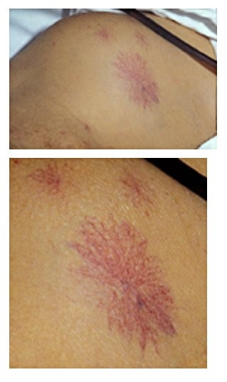
By Herbert L et al, CC BY 2.0, via Wikimedia Commons
Spider naevi may be an indication of underlying disease, particularly alcoholic cirrhosis of the liver, but can also occur in healthy individuals, especially pregnant women, or in response to pharmacological agents. Although the pathogenesis is unknown, it seems likely that they are a manifestation of disturbed circulating sex hormone activity. This may cause an elevated estradiol/free testosterone ratio. Alternatively, particularly in alcoholic cirrhosis, they may result from serum vascular growth factors in combination with alcohol, leading to neovascularisation, especially in younger patients in whom they are more common2 . Substance P may also be involved in patients with non-alcoholic cirrhosis3 .
Continue reading below
Epidemiology4
Multiple spider naevi are more indicative of underlying disease.
They have been reported in 33% of healthy children. They show an equal sex incidence.
Spider naevi may also develop in pregnancy, and were noted in 60% of pregnant women in one study.
They are more prevalent in women taking combined hormonal contraception, and disappear when this is stopped.
Just one, or a few spiders, are more common in women.
There is probably no racial difference in prevalence but they are less obvious on darker skin.
Presentation
Spider naevi are usually asymptomatic. Presentation is either due to possible concern about the aetiology or, especially if they are on the face, because of cosmetic effect. Rarely, they may bleed with minor trauma.
They are small lesions with a number of tiny blood vessels radiating from them to resemble the legs of a spider. The centre is about 1 mm in diameter and the whole lesion about 5-10 mm across. Firm pressure with a glass slide or something small, such as the end of an opened paper clip, will cause it to blanch and when the pressure is released it will rapidly refill from the central arteriole.
Lesions most frequently occur on the face, below the eyes, and over the cheekbones. Other common sites include the hands, forearms and ears. Spider naevi may occur on the trunk, or on the scalp, neck, arms and hands. Almost all spider naevi occur on the upper part of the body and only 1% below the umbilicus. Traditional teaching is that they are restricted to areas drained by the superior vena cava but this is disputed by William Bean, one of the great founders of hepatology5 . Although healthy individuals may have one or more lesions, the presence of large numbers suggests underlying disease. Look for other stigmata of liver disease.
Ask about drugs, as these may cause liver damage. Ask about alcohol consumption.
Continue reading below
Differential diagnosis
Spider naevi may be differentiated from other lesions with a similar appearance by the fact that they blanch with pressure, as described above, and refill again from the centre outwards.
Campbell de Morgan spots are bigger red spots without vessels radiating and they tend to occur in older people.
Insect bites may have a central punctum and should soon resolve. They may itch.
Dilated small blood vessels are seen in the condition telangiectasia. Osler-Weber-Rendu syndrome is a condition in which multiple areas of telangiectasia occur.
Haemangioma is a larger red spot, often raised.
Investigations
For most healthy young people with a single lesion, or just a few, and especially children, no investigation is required. Otherwise, assess liver function, including hepatitis markers. TFTs may also be indicated, as they can occur with thyrotoxicosis.
Continue reading below
Associated conditions
Spider naevi may be associated with any condition that results in increased circulating levels of oestrogen, including:
Pregnancy
Alcoholic cirrhosis
Hepatitic cirrhosis
Hepato-pulmonary syndrome
Management
Usually no treatment is required and many will fade spontaneously or resolve as the underlying condition improves. Spider naevi may be treated with laser therapy or electrocoagulation if desired for cosmetic reasons6 .
Prognosis
Benign lesions tend to resolve spontaneously but may take a number of years to do so. If associated with pregnancy they tend to go about six or seven months after delivery. They will also resolve some time after stopping combined oral contraception. If associated with liver disease, they may resolve if the liver disease improves.
Epilogue
Although Erasmus Wilson made the classic description of spider naevi, it was William Bean who studied them extensively and who noted the association with cirrhosis, especially of the alcoholic kind. He will also be remembered for his poetry on the subject of alcoholic cirrhosis, spider naevi and palmar erythema and it is worthy of repetition7 :
An older Miss Muffett
Decided to rough it
And lived upon whisky and gin.
Red hands and a spider
Developed outside her -
Such are the wages of sin.
Further reading and references
- Singh S, Sahoo AK, Ramam M, et al; Mucocutaneous spider angiomas in an adolescent with chronic liver disease. Arch Dis Child. 2018 Dec;103(12):1145. doi: 10.1136/archdischild-2017-314406. Epub 2018 Jan 3.
- Gu Y, Li K, Wu X, et al; Multiple spider angiomas in a patient with chronic hepatic graft-versus-host disease. Chin Med J (Engl). 2020 Mar 20;(6):749-750. doi: 10.1097/CM9.0000000000000707.
- Hadley, R; The Life and Works of Sir William James Erasmus Wilson, 1809-1894, Cambridge University Press, 1959.
- Li CP, Lee FY, Hwang SJ, et al; Spider angiomas in patients with liver cirrhosis: role of vascular endothelial growth factor and basic fibroblast growth factor. World J Gastroenterol. 2003 Dec;9(12):2832-5.
- Wan Y, Meng F, Wu N, et al; Substance P increases liver fibrosis by differential changes in senescence of cholangiocytes and hepatic stellate cells. Hepatology. 2017 Aug;66(2):528-541. doi: 10.1002/hep.29138. Epub 2017 Jun 19.
- Samant H, Kothadia JP; Spider Angioma
- Reuben A; Along came a spider; Hepatology 2002 Mar;35(3):735-6.1
- Erceg A, Greebe RJ, Bovenschen HJ, et al; A comparative study of pulsed 532-nm potassium titanyl phosphate laser and electrocoagulation in the treatment of spider nevi. Dermatol Surg. 2010 May;36(5):630-5. doi: 10.1111/j.1524-4725.2010.01517.x. Epub 2010 Apr 2.
- Bean WB; Vascular Spiders and Related Lesions of the Skin, Circulation, VIII, 1953.
Article History
The information on this page is written and peer reviewed by qualified clinicians.
Next review due: 20 Jul 2026
21 Jul 2021 | Latest version

Feeling unwell?
Assess your symptoms online for free