Lipoma
Peer reviewed by Dr Laurence KnottLast updated by Dr Colin Tidy, MRCGPLast updated 16 Mar 2021
Meets Patient’s editorial guidelines
Medical Professionals
Professional Reference articles are designed for health professionals to use. They are written by UK doctors and based on research evidence, UK and European Guidelines. You may find the Lumps article more useful, or one of our other health articles.
In this article:
Lipomas (lipomata) are slow-growing, benign, adipose tumours that are most often found in the subcutaneous tissues. They may also be found in deeper tissues such as the intermuscular septa, the abdominal organs, the oral cavity, the internal auditory canal, the cerebellopontine angle and the thorax. Most lipomas are asymptomatic, can be diagnosed with clinical examination and do not require treatment.
Continue reading below
Epidemiology1
Lipomas may be seen in all age groups, but usually first appear between 40 and 60 years of age.
Solitary lipomas are more common in women. Multiples - referred to as lipomatosis - are more common in men.
They have an estimated annual incidence of one per 1,000 of the population.
Congenital lipomas have been observed in children2 .
Variants
Familial multiple lipomatosis
This is an autosomal dominant condition found most frequently in men. It is characterised by widespread symmetrical lipomas appearing most often over the extremities and trunk3 .
Gardner's syndrome
Lipomatosis may also be associated with Gardner's syndrome, an autosomal dominant condition involving intestinal polyposis, cysts and osteomas4 .
Dercum's disease
This is also known as adiposis dolorosa. It is rare and characterised by the presence of irregular painful lipomas, most often found on the trunk, shoulders, arms, forearms and legs. It is at least five times more common in women than it is in men and is often found in middle age. It also has asthenia and psychiatric disturbances as other prominent features5 .
Madelung's disease
This is also known as benign symmetric lipomatosis. It is lipomatosis of the head, neck, shoulders and proximal upper extremities. Madelung's disease, often presents in men who drink excess alcohol, who may present with the characteristic 'horse collar' cervical appearance. Rarely, these patients experience swallowing difficulties, respiratory obstruction and even sudden death6 .
Post-traumatic lipomas
Lipomas have been reported that have developed following blunt trauma. It is thought that at least some of these are due to herniation of fat through torn fascia. The resulting swelling is referred to as a 'pseudo-lipoma'7 .
Continue reading below
Liposarcoma
Malignancy is rare but can be found in a lesion with the clinical appearance of a lipoma. Liposarcoma presents in a fashion similar to that of a lipoma and appears to be more common in the retroperitoneum, on the shoulders and lower extremities.
Some recommend complete excision of a lipoma to exclude a possible liposarcoma, especially in fast-growing lesions. Pre-operative radiography may be a suitable alternative in some cases8 . Both ultrasound and magnetic resonance imaging (MRI) have been used with some success to differentiate lipomas and liposarcomas but are not entirely reliable9 10 . Other modalities such as computerised tomography (CT) scanning are occasionally required11 . Alternatively, fine-needle aspiration may be used to evaluate suspicious lesions12 . National Institute for Health and Care Excellence (NICE) guidelines of 2015 advise urgent ultrasound for unexplained soft tissue lumps which are increasing in size, with two-week wait referral if ultrasound findings are suspicious or uncertain (within 48 hours for children)13 .
Tumours that have characteristics consistent with a malignant liposarcoma include those that are14 :
Greater than 5 cm in diameter.
Located in the extremities, retroperitoneally, in the groin, in the scrotum or in the abdominal wall.
Deep (beneath or fixed to superficial fascia).
Exhibiting malignant behaviour (rapid growth or invasion into nerve or bone).
In September 2020 and January 2021, NICE updated its suspected cancer recognition and referral guidance. However, no changes in these versions relate to this article13 .
Presentation
A lipoma is usually a soft or firm nodule, size 0.5-10 cm, with a smooth normal surface and skin-coloured.
While about 80% of lipomas are less than 5 cm in diameter, some can reach more than 20 cm and weigh several kilograms.
Superficial subcutaneous lipoma
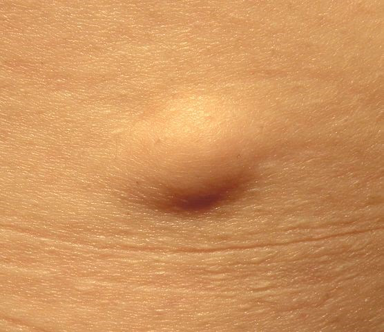
Jmarchn, CC BY-SA 3.0, via Wikimedia Commons
They are usually situated deep in the fatty layer with normal skin overlying them and should be mobile beneath the surface.
They are round or irregular in shape and may be single or multiple.
Lipomas usually present as non-painful, round, mobile masses with a characteristic soft, doughy feel.
Most lipomas are asymptomatic but they can cause pain when they compress nerves.
Continue reading below
Diagnosis
This is usually made on clinical appearance alone. Any doubt should prompt referral for imaging or specialist opinion. Ultrasound, MRI and CT scans are all used, depending on availability and index of suspicion. MRI is the imaging method of choice15 . Biopsy is not usually required if MRI is available.
Differential diagnosis
Epidermoid cyst - these may be differentiated by the punctum in their surface and also by their site in the dermis, attached to the surface.
Subcutaneous tumours.
Nodular fasciitis.
Liposarcoma.
Metastatic disease.
Nodular subcutaneous fat necrosis.
Weber-Christian panniculitis (recurring inflammation in the fat layer of the skin).
Vasculitic nodules.
Rheumatic nodules.
Infections - eg, onchocerciasis.
Haematoma.
Management
They can be left alone. The size usually plateaus after initial growth.
They may need to be removed for cosmetic reasons, because of compression of surrounding structures or if the diagnosis is uncertain.
Because lipomas generally do not infiltrate into surrounding tissue, they can usually be shelled out easily during excision.
Minimal scarring can be achieved with a technique called segmental extraction - a small stab incision followed by blind dissection of the lipoma and extraction in a segmental fashion10 .
Non-excisional treatment of lipomas includes steroid injections and liposuction10 .
Further reading and references
- Soft tissue tumors: Lipoma/benign lipomatous tumors; Atlas of Genetics and Cytogenetics in Oncology and Haematology, 2013
- Pachani AB, Shojai AR, Gautam R, et al; A giant congenital lipoma over the back. Indian J Surg. 2010 Jul;72(Suppl 1):361-2. doi: 10.1007/s12262-010-0111-7. Epub 2010 Oct 20.
- Lee CH, Spence RA, Upadhyaya M, et al; Familial multiple lipomatosis with clear autosomal dominant inheritance and onset in early adolescence. BMJ Case Rep. 2011 Feb 17;2011. pii: bcr1020103395. doi: 10.1136/bcr.10.2010.3395.
- Chatziralli IP, Papazisis L, Sergentanis TN; Incomplete Gardner's syndrome with blepharoptosis as the first symptom. Int Ophthalmol. 2013 Apr 12.
- Hansson E, Svensson H, Brorson H; Review of Dercum's disease and proposal of diagnostic criteria, diagnostic methods, classification and management. Orphanet J Rare Dis. 2012 Apr 30;7:23. doi: 10.1186/1750-1172-7-23.
- Suresh Chandran CJ, Godge YR, Oak PJ, et al; Madelung's disease with myopathy. Ann Indian Acad Neurol. 2009 Apr;12(2):131-2. doi: 10.4103/0972-2327.53086.
- Rathi NV, Dahake PT, Thakre K, et al; Traumatic pseudo-lipoma in 3-year-old child. Contemp Clin Dent. 2012 Oct;3(4):487-90. doi: 10.4103/0976-237X.107451.
- Jones AP, Lewis CJ, Dildey P, et al; Lipoma or liposarcoma? A cautionary case report. J Plast Reconstr Aesthet Surg. 2012 Jan;65(1):e11-4. doi: 10.1016/j.bjps.2011.08.004. Epub 2011 Aug 23.
- Brisson M, Kashima T, Delaney D, et al; MRI characteristics of lipoma and atypical lipomatous tumor/well-differentiated liposarcoma: retrospective comparison with histology and MDM2 gene amplification. Skeletal Radiol. 2013 May;42(5):635-47. doi: 10.1007/s00256-012-1517-z. Epub 2012 Sep 18.
- Nguyen T, Zuniga R; Skin conditions: benign nodular skin lesions. FP Essent. 2013 Apr;407:24-30.
- Weerakkody Y et al; Liposarcoma, Radiopaedia.org
- Jagtap SV, Nikumbh DB, Jagtap SS, et al; Huge dedifferentiated liposarcoma of the left thigh with a high grade fibrosarcomatous differentiation and a local recurrence. J Clin Diagn Res. 2013 Mar;7(3):553-6. doi: 10.7860/JCDR/2013/4610.2823. Epub 2013 Mar 1.
- Suspected cancer: recognition and referral; NICE guideline (2015 - last updated October 2023)
- Costea R, Vasiliu E, Zarnescu NO, et al; Large thigh liposarcoma--diagnostic and therapeutic features. J Med Life. 2011 May 15;4(2):184-8. Epub 2011 May 25.
- Abd Rabou A et al; Lipoma; Radiopaedia.org
Article History
The information on this page is written and peer reviewed by qualified clinicians.
Next review due: 15 Mar 2026
16 Mar 2021 | Latest version

Feeling unwell?
Assess your symptoms online for free