Pilar cysts
Epidermoid and sebaceous cysts
Peer reviewed by Dr Laurence KnottLast updated by Dr Colin Tidy, MRCGPLast updated 20 Nov 2021
Meets Patient’s editorial guidelines
Medical Professionals
Professional Reference articles are designed for health professionals to use. They are written by UK doctors and based on research evidence, UK and European Guidelines. You may find the Epidermoid and pilar cysts article more useful, or one of our other health articles.
In this article:
Synonyms: sebaceous cysts (a misnomer as these cysts are not of sebaceous origin), trichilemmal cysts
Continue reading below
What are epidermoid and pilar cysts?
The vast majority of epidermoid or pilar cysts are of no great consequence.
Epidermal cysts (also known as epithelial or infundibular cysts) are intradermal or subcutaneous tumours. Epidermoid cysts may occur anywhere on the body but occur most often on the face, scalp, neck, back and scrotum.
Pilar cysts (also called trichilemmal cysts) are clinically indistinguishable from epidermal cysts. They contain keratinous material, are usually multiple and there is often an autosomal dominant inheritance.
Who gets epidermoid and pilar cysts? (Epidemiology)
They are both extremely common and probably most people will have at least one over the course of a lifetime. They can often resolve spontaneously.
They are around twice as common in men as in women and most frequent in those aged in their 20s and 30s.
Simple epidermoid cysts can run in families.
Epidermoid cyst on the neck
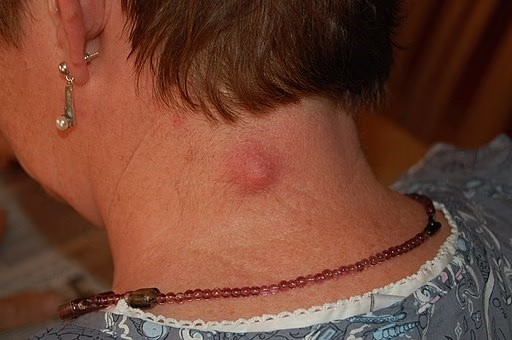
By Steven Fruitsmaak, CC BY-SA 3.0, via Wikimedia Commons
Continue reading below
Epidermoid and pilar cyst symptoms
Most often they present as a painless skin lump.
They may present with discharge of a foul cheese-like material.
If they become infected, they are red, inflamed and painful.
Lesions of the genitals can be painful during intercourse and cause problems with walking or wearing underwear. They can also interfere with micturition.
Epidermoid and pilar cysts appear as firm, round, mobile, flesh-coloured to yellow or white subcutaneous nodules of variable size.
A central pore or punctum may tether the cyst to the overlying epidermis and a thick cheesy material can sometimes be expressed.
In people with dark skin, the cysts may also be pigmented.
Sites affected by epidermoid and pilar cysts
The sites most commonly affected are, in descending order of frequency, the face, the trunk, the neck, the extremities and the scalp.
Cysts of the genitals are less common and may appear as a mass in the vulva, the clitoris, the penis, the scrotum, or the perineum. In cultures which practise female circumcision (or female genital mutilation) cysts on the vulva are common.
The ocular and oral mucosae can also be affected and cysts have been reported on the palpebral conjunctivae, on the lips, on the buccal mucosa, on and under the tongue and even on the uvula. These are not sites with hair follicles.
Cysts may occur on the extremities. Subungual cysts can cause changes in the nails, such as onycholysis and subungual hyperkeratosis, which may be mistaken for psoriasis or onychomycosis. These cysts also produce changes in the nails (such as pincer nails) erythema, oedema, tenderness and pain.
Palmoplantar lesions represent a unique subset of epidermoid cysts.
Differential diagnosis
Other causes of skin cysts.
A lipoma tends to be larger and is very soft.
A neurofibroma is hard and may be multiple.
An abscess is hot and red and may resemble an infected sebaceous cyst.
Multiple cysts in a teenager may suggest Gardner's syndrome.
Continue reading below
Investigations
Usually the diagnosis is clear and no investigations are required. In exceptional cases malignancy may be suspected, in which case excision and histology are required.
Associated diseases
Epidermoid cysts are a feature of Gardner's syndrome. Gardner's syndrome is an autosomal dominant condition comprising familial polyposis coli, cutaneous cysts and osteomas or other soft-tissue tumours. The cysts tend to occur at an earlier age than usual (presenting most often in the early teenage years). The face may be involved but the extremities tend to be affected more than the trunk.
Epidermoid and pilar cyst treatment and management
Most people with an epidermoid or pilar cyst never seek medical attention.
If a cyst is uncomplicated then no treatment is usually advisable. The cyst may disappear spontaneously, leaving no trace. Even the most skilful excision will leave a permanent scar.
If the cyst is red and hot it is probably infected. An antibiotic effective against staphylococci should be used - eg, flucloxacillin. Infection may be mixed and, in lesions of the scalp and anogenital area, anaerobic flora are more likely.
If the cyst has ruptured, the contents can be expressed. However, the cyst may well re-form.
An inflamed but uninfected cyst may respond to intralesional injection of steroid but it is not easy to tell if an inflamed cyst is infected or not and this is not usually recommended.
If the cyst is troublesome or if the patient, after counselling, is eager to have it removed then the entire cyst should be excised as a surgical procedure.
Excision of an epidermoid or pilar cyst
See also separate article Minor Surgery in Primary Care.
The usual preliminaries of informed consent are assumed.
Information includes the warning that the cyst may recur if all the wall is not removed.
Appropriate aseptic precautions should be taken.
Local anaesthetic such as 1% lidocaine with adrenaline (epinephrine) is used (note contra-indications to use of adrenaline (epinephrine) in certain areas). The area is infiltrated with local anaesthetic, being careful not to puncture the cyst.
Make a careful and superficial incision over the cyst avoiding rupture of the cyst. Care should be taken to remove all the cyst wall if the cyst is ruptured.
Toothed forceps can be used to grip the skin - a blunt dissection of adhesions allows mobilisation of the cyst and removal. A Volkmann spoon or scissors are useful for the blunt dissection.
If the cyst is large and leaves a significant defect then a resorbable subcutaneous suture should be used to close a potential space for haematoma to form.
The diagnosis of an epidermoid or pilar cyst is usually obvious. However, the excised tissue should be submitted for histology.
Complications with epidermoid or pilar cysts
Anxiety or cosmetic embarrassment.
Infection.
Malignant change is very rare (causes rapid growth, friability and bleeding).
Genital and umbilical lesions sometimes extend into the pelvis or below the midline fascia. This may not be apparent until they are being removed.
Very rarely, lesions over the skull can have an intracranial or intraosseous connection. Inappropriate management may result in cerebrospinal fluid leakage and complications (meningitis and death). Preliminary CT or MRI scan may be of value1 . Features suggestive of intracranial or intraosseous extension include the following:
Present since birth or appearance in early childhood (although small lesions may remain unnoticed until adulthood).
Bruits, pulsation or fluctuation in size with straining or crying.
Fixation to underlying tissue, fluid-filled consistency, or ability to transilluminate.
Location along the nasal, forehead or scalp midline, or along cranial suture lines.
Dimple or unusual overlying hair growth pattern.
History of cranial trauma or surgery.
Family history of neural developmental anomalies, neurological symptoms or history of meningitis.
Intracranial epidermoid cysts are rare, histologically benign, slow-growing, congenital neoplasms of the central nervous system that may arise from retained ectodermal implants2 .
Prognosis
They will usually grow slowly and only need removal if causing symptoms. They tend to recur if incised rather than excised.
Further reading and references
- Al Aboud DM, Yarrarapu SNS, Patel BC; Pilar Cyst. StatPearls, August 2021.
- Trichilemmal cyst; DermNet NZ.
- Pilar cyst; Primary Care Dermatology Society.
- Sorenson EP, Powel JE, Rozzelle CJ, et al; Scalp dermoids: a review of their anatomy, diagnosis, and treatment. Childs Nerv Syst. 2013 Mar;29(3):375-80. doi: 10.1007/s00381-012-1946-y. Epub 2012 Nov 21.
- Vellutini EA, de Oliveira MF, Ribeiro AP, et al; Malignant transformation of intracranial epidermoid cyst. Br J Neurosurg. 2014 Aug;28(4):507-9. doi: 10.3109/02688697.2013.869552. Epub 2013 Dec 18.
Article History
The information on this page is written and peer reviewed by qualified clinicians.
Next review due: 19 Nov 2026
20 Nov 2021 | Latest version

Feeling unwell?
Assess your symptoms online for free