Iron-deficiency anaemia
Peer reviewed by Dr Laurence KnottLast updated by Dr Colin Tidy, MRCGPLast updated 28 Jul 2020
Meets Patient’s editorial guidelines
Medical Professionals
Professional Reference articles are designed for health professionals to use. They are written by UK doctors and based on research evidence, UK and European Guidelines. You may find one of our health articles more useful.
In this article:
There are separate Childhood Anaemia and Anaemia in Pregnancy articles.
Iron-deficiency anaemia (IDA) occurs when the body has insufficient iron to support red blood cell production.
The World Health Organization defines anaemia as:1
Haemoglobin (Hb) <130 g/L in men aged 15 years or older.
Hb <120 g/dL in non-pregnant women aged 15 years or older.
Failure to investigate IDA appropriately in primary care can cause significant delay in final diagnosis, with associated morbidity.
Continue reading below
Epidemiology
Iron deficiency is the most common cause of anaemia worldwide, affecting around 500 million people.2
In the developed world, 2-5% of adult men and postmenopausal women have IDA.
Iron deficiency and IDA are common in young children.3
Iron deficiency is the most common cause of anaemia in pregnancy worldwide; severe anaemia can have very serious consequences for mothers and babies.4
Premenopausal women have a higher incidence of IDA because of heavy menstrual blood losses and pregnancy.
Aetiology2
Causes of iron deficiency may be classified as those due to:
Excessive blood loss
Blood loss from the gastrointestinal (GI) tract is the most common cause of IDA in adult men and postmenopausal women. 5
Blood loss due to menorrhagia is the most common cause of iron deficiency in pre-menopausal women.
In tropical countries, infestation of the gut may cause IDA, especially with hookworm and schistosomiasis.
Common causes of blood loss include:
Non-steroidal anti-inflammatory drug (NSAID) use.
Angiodysplasia.
Other causes include:
Other GI tract malignancies.
Bleeding oesophageal varices.
Inflammatory bowel disease.
Haemorrhoids.
Oesophagitis and gastro-oesophageal reflux disease.
Postpartum haemorrhage.
Recurrent epistaxis.
Malignancy of the renal tract.
After major surgery or major trauma, if replacement has been inadequate.
After blood donation.
Dietary inadequacy
Dietary iron deficiency is fairly uncommon.
Meat tends to be more rich than vegetables in iron and so vegetarians are at greater risk. However, green vegetables are a good source of iron and a proper vegetarian diet should not lead to deficiency.
Growing children and elderly people with iron-poor diets may become deficient.
Failure of iron absorption
Not only does the diet have to contain adequate amounts of iron but the iron has to be in a form that can be absorbed.
Iron can be absorbed in the ferrous state much more readily than in the ferric state.
Factors affecting iron absorption:
Some drugs can bind to iron and prevent absorption. Tetracyclines and quinolones chelate with iron so that neither the antibiotic nor the iron is absorbed.
Antacids and proton pump inhibitors may also impair absorption by raising gastric pH.2
Phytate (found in wholegrain cereals, nuts, seeds and legumes), polyphenols (found in tea and coffee) and calcium (in dairy products) impair iron absorption.
Iron absorption can be increased in a diet rich in fish, red meat and white meat.
Vitamin C may enhance iron absorption. Patients can be encouraged to drink a glass of orange juice with their iron tablets.
Malabsorption conditions such as coeliac disease (usually accompanied by folate deficiency).
It may occur after partial or total gastrectomy, more commonly with increased number of postoperative years.
Helicobacter pylori colonisation appears to impair iron uptake and increase iron loss.
Excessive requirements for iron
Times of high demand for iron should be met by greater absorption from the diet.
If the diet is not adequate, an intake that would otherwise be sufficient becomes inadequate - for example in:
Times of rapid growth in children.
Pregnancy, especially with twins.
Exfoliative skin disease.
Continue reading below
Presentation
IDA is often an incidental finding rather than a presenting feature. Chronic, slow blood loss can lead to compensation by the body and little in the way of symptoms. If symptoms occur, they can include:2
Fatigue.
Shortness of breath on exertion.
Palpitations.
Sore tongue and taste disturbance.
Changes in the hair/hair loss.
Pruritus.
Headache.
Tinnitus.
Angina, which can occur if there is pre-existing coronary heart disease.
Very rarely, dysphagia due to an oesophageal web with chronic iron deficiency. This is the Plummer-Vinson syndrome and there is an association with oesophageal carcinoma.
Symptoms of severe anaemia (usually not occurring until Hb is <7 g/dL) include: shortness of breath at rest, angina and ankle swelling. These symptoms may occur at higher Hb levels if there is co-existing cardiorespiratory disease.2
History
Cover the following points looking for potential causes:
Current/recent diet - poor iron intake or impaired absorption.
Drug history - NSAIDs, selective serotonin reuptake inhibitors (SSRIs), clopidogrel, and corticosteroids could all be a potential cause.
Any overt bleeding seen by the patient - eg, nosebleeds, rectal bleeding.
History of recent blood donation.
Menstrual history in women.
History of recent illness: could suggest GI bleeding - eg, weight loss, change in bowel habit, dyspepsia.
History of previous GI disease or surgery.
Travel history (eg, hookworm infestation) if there has been recent travel to the tropics.
Family history, including inherited haematological disorders such as thalassaemia; bleeding disorders and telangiectasia; IDA, as may indicate a potential inherited disorder of iron absorption.
Continue reading below
Examination
Examine the abdomen for abdominal masses, organomegaly, lymphadenopathy and any other features of intra-abdominal disease.
Rectal examination is seldom helpful unless there is rectal bleeding or tenesmus. Postpone until colonoscopy.
If menorrhagia is thought to be the cause: perform a vaginal/bimanual examination, examine the cervix and perform a cervical smear and swabs as appropriate (see separate article Menorrhagia for further details).
Signs
Pallor (best seen in the mucosa of the tongue and mouth, especially in people with dark skin).
Koilonychia (spoon-shaped nails with longitudinal ridging).
Angular cheilitis (ulceration at the corners of the mouth).
Atrophic glossitis.
In marked anaemia, there may be tachycardia, a flow murmur, cardiac enlargement, ankle oedema and heart failure.
KOILONYCHIA
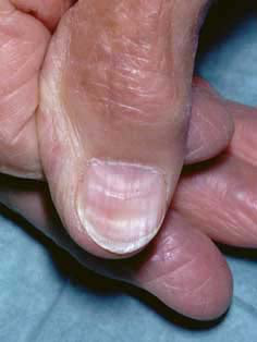
Note the spoon-shaped nail of koilonychia with the longitudinal ridges.
Other signs that may be seen include:
Stigmata of chronic liver disease, perhaps cirrhosis.
Multiple telangiectasias, which may be a feature of hereditary haemorrhagic telangiectasia, also known as Osler-Weber-Rendu syndrome.
Pigmentation of the lips and oral mucosa, which may suggest Peutz-Jeghers syndrome.
Confirming the diagnosis
IDA can be diagnosed in most cases by FBC and serum ferritin level.6
FBC: shows a hypochromic microcytic anaemia (although there may be a mixed picture with co-existent B12 or folate deficiency):
Hypochromia means that there is as a low mean corpuscular haemoglobin (MCH).
Microcytosis means that there is as a low mean corpuscular volume (MCV).
Remember that a haemoglobinopathy will also cause a hypochromic microcytic anaemia.
Serum ferritin: should be measured to confirm iron deficiency (except during pregnancy):
This correlates with total body iron stores.
However, ferritin levels can be raised if infection or inflammation is present, even if iron stores are low.
A cut-off ferritin level varies between 12-15 mcg/L to confirm iron deficiency.
If there is co-existing chronic inflammatory disease, the clinician should consider seeking specialist advice about other measures of iron status.
Blood film: anisocytosis (variation in size between red blood cells) and poikilocytosis (abnormally shaped red blood cells) can be seen.
Differential diagnosis
Other causes of microcytic anaemia including:
Investigations
Urine should be tested in all patients.
All male and postmenopausal females should be considered for upper and/or lower GI investigations. Endoscopy or CT scan are acceptable. Endoscopy is preferred.
All patients should be screened for coeliac disease.
If oesophagogastroduodenoscopy (OGD) is the initial investigation, lower GI endoscopy should also be performed unless advanced gastric cancer or coeliac disease is found. Even when coeliac disease has been found, consider colonoscopy if the patient is aged over 50 years or has a significant family history of colonic carcinoma.
If there are symptoms suggesting small bowel disease or if normal iron levels cannot be restored (or maintained) by iron therapy, direct visualisation of the small bowel may be necessary.
If the patient has recurrent IDA and normal OGD and colonoscopy, testing for (and subsequent eradication of) H. pylori is advised.
Pre-menopausal women should have endoscopy if they are over 50 years old, or there are suggestive GI symptoms or there is a strong family history of colorectal cancer.
Upper and lower GI investigations are recommended for patients aged over 50 years, who have had a gastrectomy.
Only postmenopausal women and men aged over 50 years should have GI investigations of iron deficiency without anaemia.
NB: the appropriateness of investigating patients who are frail and/or have significant comorbidity needs to be considered on an individual basis. The severity and nature of the anaemia should be weighed against the risk of bowel preparation and whether the patient is fit enough to withstand treatment (if a colorectal cause was found).
Who should be referred to secondary care?2 7
Editor’s note
Dr Sarah Jarvis, 11th February 2021
In September 2020 and, more recently, January 2021, the National Institute for Health and Care Excellence (NICE) issued updated guidance on suspected cancer recognition and referral. There are, however, no changes relating to recognition of, and referral for, IDA.
Urgent referral using a suspected cancer pathway for an appointment within two weeks if they are aged 60 years or over, or aged under 50 years and present with rectal bleeding.
Refer to gastroenterology:
All men and postmenopausal women with IDA unless they have overt non-gastrointestinal bleeding. Men with Hb less than 120 g/L and postmenopausal women with an Hb level less than 100 g/L should be investigated more urgently, as lower levels of Hb suggest more serious disease.
All people aged 50 years or over with marked anaemia, or a significant family history of colorectal carcinoma, even if coeliac disease is found.
Premenopausal women if they are aged under 50 years and have colonic symptoms, a strong family history (two affected first-degree relatives or just one first-degree relative affected before the age of 50 years) of gastrointestinal cancer, or persistent IDA despite treatment.
Refer women to gynaecology if:
Menorrhagia is unresponsive to medical management.
Postmenopausal bleeding (suspected cancer pathway for an appointment within two weeks).
Pregnant and has significant symptoms and/or severe anaemia (Hb less than 70 g/L), or late gestation (over 34 weeks), or if there is failure to respond to a trial of oral iron.
Also refer people:
If coeliac serology is positive - refer to gastroenterology.
If they have profound anaemia with signs of heart failure.
If they are unable to tolerate, or are not responding to, oral iron treatment.
Who have initially responded to iron treatment but develop anaemia again without an obvious underlying cause.
When the type of anaemia is in doubt.
When further haematological investigations, such as bone marrow examination or an investigation of bleeding state, cannot be carried out in primary care.
Treatment
Treatment for the iron deficiency should be started before the results of the investigations.
Iron salts should be given by mouth unless there are good reasons for using another route. Ferrous salts show only marginal differences between one another in efficiency of absorption of iron.8
When the underlying cause is known, this should be managed appropriately.
Iron-rich foods include dark green vegetables, meat, apricots, prunes, raisins and iron-fortified bread.
Consider referral to a dietician if diet is thought to be the cause.
Parenteral iron therapy8
Parenteral iron is generally reserved for use when oral therapy is unsuccessful because the patient cannot tolerate oral iron or does not take it reliably, if there is continuing blood loss, or in malabsorption.
With the exception of patients with severe renal failure receiving haemodialysis, parenteral iron does not produce a faster haemoglobin response than oral iron provided that the oral iron preparation is taken reliably and is absorbed adequately.
Iron can be administered parenterally as iron dextran, iron sucrose, ferric carboxymaltose, or ferric derisomaltose.
Many patients with chronic renal failure who are receiving haemodialysis (and some who are receiving peritoneal dialysis) also require iron by the intravenous route on a regular basis.
Side-effects to iron supplementation
Common side-effects usually reduce with time and include:
Constipation
Black stools
Diarrhoea
Heartburn
Nausea
Abdominal/epigastric pain
Intravenous iron therapy may cause hypophosphataemia.
Monitoring response to treatment
FBC should be checked 2-4 weeks after treatment has started.2
If there is response to treatment
Check FBC again in 2-4 months to ensure Hb levels have returned to normal.
Once Hb levels are normal, continue treatment for three months.
Re-check FBC every three months for one year.
Re-check again after a further year.
If Hb or red cell indices drop below normal, give additional iron.
If there is inadequate response to treatment
Assess compliance; is the iron supplement tolerated?
If there are problems with compliance:
Consider prescribing a laxative if constipation is experienced.
Advise the patient to take iron with or after meals.
Reassure the patient that black stools are normal and harmless.
Reduce the frequency of the iron supplement to one or two times per day.
Try a different formulation with a lower content of elemental iron - eg, ferrous gluconate.
If oral supplements are still not tolerated, consider asking for specialist advice. If the patient is unable to tolerate oral iron or there is malabsorption, parenteral iron can be given. This is not recommended in primary care.2
Transfusion is occasionally necessary and is reserved for those at risk of cardiovascular instability.
If there is an increase of less than 2 g/dL in the Hb level after 2-4 weeks, refer for specialist assessment and advice.
Further reading and references
- NDR (Nutrition and Diet Resources) UK
- Investigation and Management of the Adult Patient with anaemia; Guideline Audit and Implementation Network, 2015
- Haemoglobin concentrations for the diagnosis of anaemia and assessment of severity; World Health Organization, 2011
- Anaemia - iron deficiency; NICE CKS, September 2018 (UK access only)
- Wang B, Zhan S, Gong T, et al; Iron therapy for improving psychomotor development and cognitive function in children under the age of three with iron deficiency anaemia. Cochrane Database Syst Rev. 2013 Jun 6;6:CD001444. doi: 10.1002/14651858.CD001444.pub2.
- Reveiz L, Gyte GM, Cuervo LG, et al; Treatments for iron-deficiency anaemia in pregnancy. Cochrane Database Syst Rev. 2011 Oct 5;(10):CD003094.
- Ageing and Life Course; World Health Organization
- Pasricha SR, Flecknoe-Brown SC, Allen KJ, et al; Diagnosis and management of iron deficiency anaemia: a clinical update. Med J Aust. 2010 Nov 1;193(9):525-32.
- Suspected cancer: recognition and referral; NICE guideline (2015 - last updated October 2023)
- British National Formulary (BNF); NICE Evidence Services (UK access only)
Article History
The information on this page is written and peer reviewed by qualified clinicians.
Next review due: 27 Jul 2025
28 Jul 2020 | Latest version

Feeling unwell?
Assess your symptoms online for free