
The respiratory system
Peer reviewed by Prof Cathy Jackson, MRCGPLast updated by Dr Colin Tidy, MRCGPLast updated 31 May 2018
The lungs are divided into different parts which are called lobes. The right lung has three lobes called upper, middle and lower lobes. The left lung only has two lobes, the upper and lower.
In this article:
Continue reading below
Where are the lungs found?
The lungs are found in the chest on the right and left side. At the front they extend from just above the collarbone (clavicle) at the top of the chest to about the sixth rib down. At the back of the chest the lungs finish around the tenth rib. The protective linings which cover the lungs (pleura) continue down to the twelfth rib. From front to back the lungs fill the rib cage but are separated by the heart, which lies in between them.
Chest and lungs fully labelled
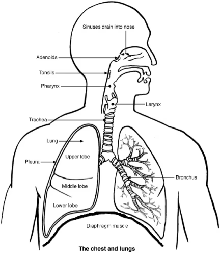
The air that we breathe in enters the nose or mouth, flows through the throat (pharynx) and voice box (larynx) and enters the windpipe (trachea). The trachea divides into two hollow tubes called bronchi. The right main bronchus (bronchus is the word for one of the bronchi) supplies the right lung; the left main bronchus supplies the left lung. These bronchi then go on to divide into smaller bronchi. The small bronchi divide into smaller and smaller hollow tubes which are called bronchioles - the smallest air tubes in the lungs. The medical term for all the air tubes from the nose and mouth down to the bronchioles is 'the respiratory tract'. The lower respiratory tract is from the larynx.
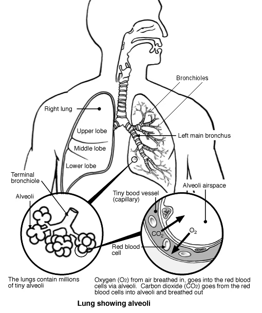
At the end of the smallest bronchioles are tiny air sacs called alveoli. Alveoli are lined by a very thin layer of cells. They also have an excellent blood supply. The tiny alveoli are the place where oxygen enters the blood and where carbon dioxide (CO2) leaves the blood.
Alveoli detail
Patient picks for Healthcare


What do the lungs do?
The lungs' main function is to help oxygen from the air we breathe enter the red cells in the blood. Red blood cells then carry oxygen around the body to be used in the cells found in our body. The lungs also help the body to get rid of CO2 gas when we breathe out. There are a number of other jobs carried out by the lungs that include:
Changing the pH of blood (whether the blood is more acid or alkali) by increasing or decreasing the amount of CO2 in the body.
Filtering out small gas bubbles that may occur in the bloodstream.
Converting a chemical in the blood called angiotensin I to angiotensin II. These chemicals are important in the control of blood pressure.
Continue reading below
How do the lungs and breathing work?
Breathing in is called inhalation. The most important muscle of inhalation is the diaphragm. Found beneath the lungs, the diaphragm is a dome-shaped muscle. When this muscle gets tighter (contracts), it flattens and the lungs increase in size. This sucks air down into your lungs.
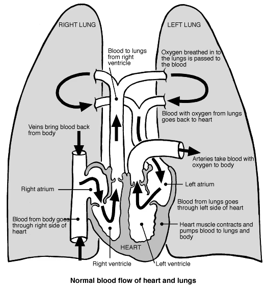
Some of the oxygen in the air can then be transferred into your bloodstream. Some of the carbon dioxide in your blood is transferred into the air that is in your lungs. This controls the levels of oxygen and carbon dioxide in your bloodstream. See also the separate leaflet called The Heart and Blood Vessels for more information on how blood is pumped to the lungs and to the rest of your body.
Breathing out (exhalation) is the opposite of inhalation. The diaphragm and other chest muscles relax. This makes the lungs decrease in size so that air is pushed back out of your lungs and out through your mouth or nose.
The basic rhythm of breathing is controlled by the brain. Part of the brain called the brainstem has a special area dedicated to maintaining your breathing pattern. Nerve impulses from the brainstem control the contractions of your diaphragm and the other muscles of breathing. This is all done without thinking. However, other parts of the brain can temporarily overrule the brainstem. This is how we are able consciously to hold our breath or change our pattern of breathing.
While the brain controls the basic rhythm of breathing, it also receives information from sensors in the body. These sensors are nerve cells and provide information that influences the rate and depth of breathing. The main sensors monitor levels of CO2 in the blood.
When the level of CO2 rises, the sensors send electrical impulses to the brain. These impulses cause the brain to send more electrical signals to the muscles of breathing. Breathing then gets deeper and faster and more CO2 is breathed out (exhaled). The blood level of CO2 then decreases back to the normal level.
Heart and lungs - normal blood flow
Some disorders of the respiratory tract, lung and chest
Continue reading below
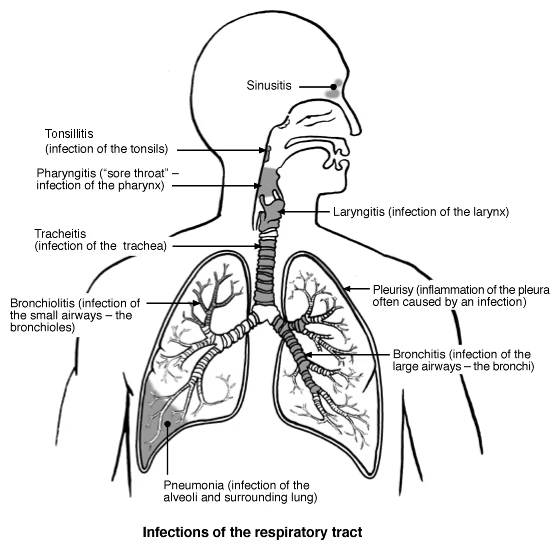
Some infections of the respiratory tract
The diagram below shows in which part of the respiratory tract some of the infections are located:
Respiratory tract infections
Article History
The information on this page is written and peer reviewed by qualified clinicians.
31 May 2018 | Latest version

Feeling unwell?
Assess your symptoms online for free