Spontaneous pneumothorax - complications after surgery - now have hydrothorax - very confused
Posted , 3 users are following.
Late September while interstate I was diagnosed with a spontaneous fully collapsed left lung with severe shift. A test tube was inserted and over several days air was extracted allowing the lung to inflate to 3/4 of its original size. Experienced localized pain a week later in the middle directly under breast. An xray was taken and a small amount of fluid was found in that area. They did not know what it was and just said it was an infection. I was given anti biotics for two weeks. The pain went away shortly after taken the medication. Two weeks later I returned to the hospital for a review. The only issue noted by the doctors after having an xray was that the lung was still not fully expanded (only 3/4) and that the air left in the lung which was supposed to have been processed into the body had not done so. There was no note at that time of any other air being present in the lung cavity. A few weeks later I returned home to Melbourne and visited my gp as I was still feeling breathless and generally unwell/fatigued. My gp gave me a referral to the emergency department . Xrays from the ED showed a large hydrothorax. The doctors say I need surgery to remove it. They have told me that my lung never collapsed, even though they had a disc with all the xrays taken at the other hospital and that it was the hydrothorax that caused my symptoms initially. They did not have the images of the xrays taken interstate at that time. I only have a letter from them that they wrote before they got ahold of the xray images from the interstate hospital. have been sent home. I'm really confused because I do not understand how a hydrothorax could have been present at the time of the first operation as my symptoms improved and the hydrothorax was not noted at all in the first xrays. It was also not found when I returned for a review after the first operation. Any help would be much appreciated.
The preliminary report says -
Reason for exam - 59f large hydrothorax, recent spontaneous pneumothorax (managed Tasmania), ?ruptured pneumoatocele (request per cardiothoracix)
Technique - CT chest no prior imaging available
Findings - Large round cystic mass occupying majority of the left upper lobe, demonstrates thin walls and an internal air-fluid llevel (fuid density approx HU 10). There is adjacent atelectasis and compression of the left upper lobe, with mild mediastinal shift to the right. no convincing pneumothorax or evidence of rupture is identified.
Normal appearence of background lung parenchyma. No medastinal or axillary lymphadenopathy. Normal appearence and enhancement of the mediastinal vasculature.
Partially imaged upper abdominal viscera appear normal. No osseous lessions.
Conclusion - Large round cystic mass in the left upper lobe is favoured to represent a pneumatocele - it contains fluid and exerts mild mass effect on the adjacent structures. No evidence of rupture or pneumothorax. 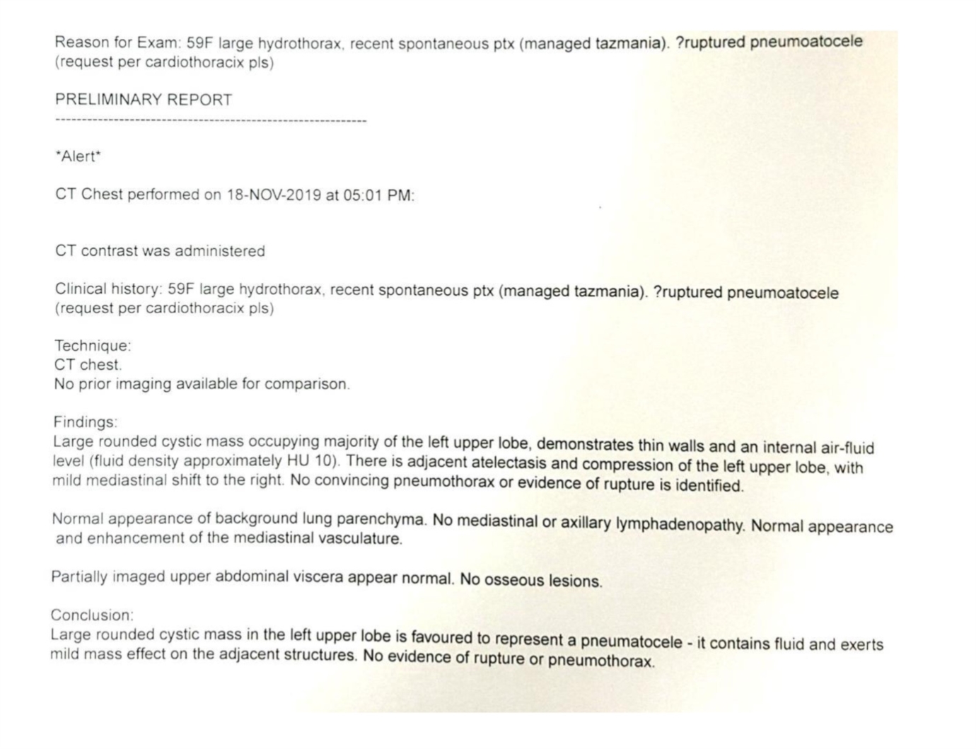
0 likes, 1 reply
amkoffee dayle12721
Posted
I can not speak of everything going on with your health but I'm sure your very frightened by it all. The one thing I have learned in the last several years is that doctors are not infallible. They make mistakes. I'm not trying to be disparaging towards doctors but they are human and they make mistakes. It is for this reason that I urge you to seek a second or third opinion.