Gyrate atrophy of the choroid and retina
Last updated 18 Aug 2020
Meets Patient’s editorial guidelines
- DownloadDownload
- Share
- Language
- Discussion
- Ask Patient
- Audio Version
- Add to preferred sources on Google
Gyrate atrophy of the choroid and retina, which is often shortened to gyrate atrophy, is an inherited disorder characterised by progressive vision loss. People with this disorder have an ongoing loss of cells (atrophy) in the retina, which is the specialized light-sensitive tissue that lines the back of the eye, and in a nearby tissue layer called the choroid. During childhood, they begin experiencing nearsightedness (myopia), difficulty seeing in low light (night blindness), and loss of side (peripheral) vision. Over time, their field of vision continues to narrow, resulting in tunnel vision. Many people with gyrate atrophy also develop clouding of the lens of the eyes (cataracts). These progressive vision changes lead to blindness by about the age of 50.
Video picks for Genetic conditionsVideo library
Description
Gyrate atrophy of the choroid and retina, which is often shortened to gyrate atrophy, is an inherited disorder characterised by progressive vision loss. People with this disorder have an ongoing loss of cells (atrophy) in the retina, which is the specialized light-sensitive tissue that lines the back of the eye, and in a nearby tissue layer called the choroid. During childhood, they begin experiencing nearsightedness (myopia), difficulty seeing in low light (night blindness), and loss of side (peripheral) vision. Over time, their field of vision continues to narrow, resulting in tunnel vision. Many people with gyrate atrophy also develop clouding of the lens of the eyes (cataracts). These progressive vision changes lead to blindness by about the age of 50.
Most people with gyrate atrophy have no symptoms other than vision loss, but some have additional features of the disorder. Occasionally, newborns with gyrate atrophy develop excess ammonia in the blood (hyperammonemia), which may lead to poor feeding, vomiting, seizures, or coma. Neonatal hyperammonemia associated with gyrate atrophy generally responds quickly to treatment and does not recur after the newborn period.
Gyrate atrophy usually does not affect intelligence; however, abnormalities may be observed in brain imaging or other neurological testing. In some cases, mild to moderate intellectual disability is associated with gyrate atrophy.
Gyrate atrophy may also cause disturbances in the nerves connecting the brain and spinal cord to muscles and sensory cells (peripheral nervous system). In some people with the disorder these abnormalities lead to numbness, tingling, or pain in the hands or feet, while in others they are detectable only by electrical testing of the nerve impulses.
In some people with gyrate atrophy, a particular type of muscle fibers (type II fibers) break down over time. While this muscle abnormality usually causes no symptoms, it may result in mild weakness.
Inheritance patterns
The source lists the following possible inheritance patterns: Autosomal recessive. More than one pattern can apply in different cases.
Related genes
Other names
Gyrate atrophy
HOGA
Hyperornithinemia with gyrate atrophy of choroid and retina
OAT deficiency
OKT deficiency
Ornithine aminotransferase deficiency
Ornithine keto acid aminotransferase deficiency
Ornithine-delta-aminotransferase deficiency
Ornithinemia with gyrate atrophy
Patient picks for Genetic conditions
Preventative health
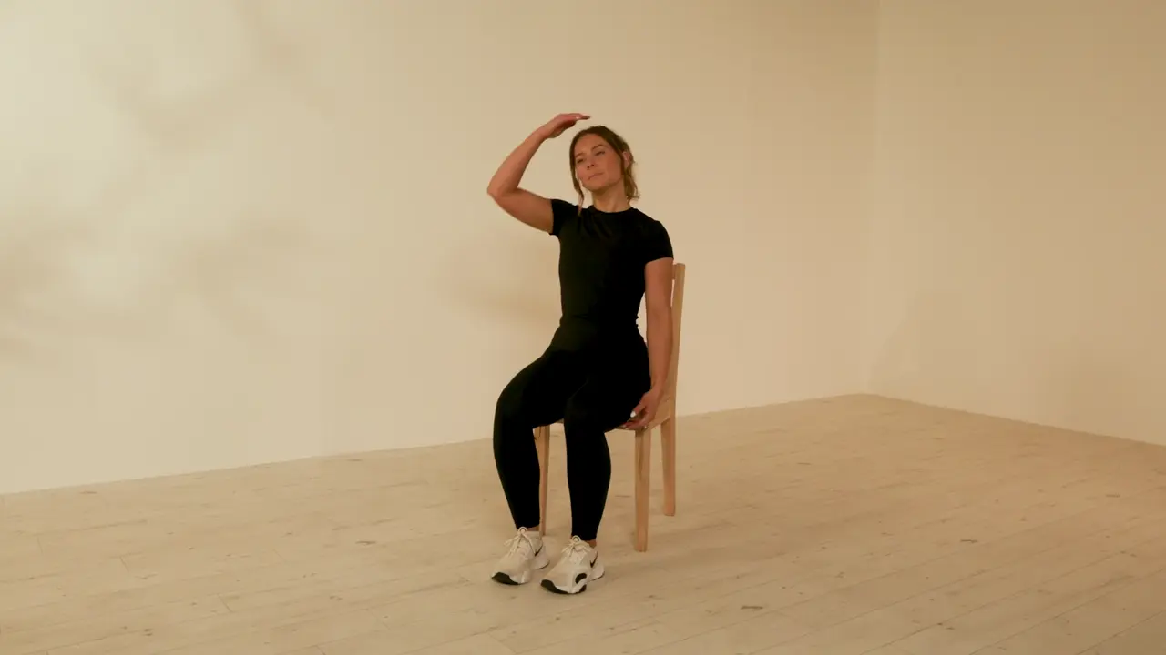
Seated Neck Stretch
This is a simple exercise aimed at stretching the muscles of the neck and upper back.
Podcasts
Beyond human
Have you’ve ever felt transformed after a walk by the sea or in the woods? Have you’ve ever found yourself reaching for the biscuit tin when anxious rather than hungry, or noticed how your pet knows when you are feeling low? This podcast is for you. Clare and Aileen take you on a tour of some of those relationships we often don’t think about but matter deeply. From pets and nature to time, food and even life and the divine, these relationships have the ability to shape our lives, comfort us, challenge us and sometimes reveal parts of ourselves we didn’t know were there.
by Thomas Andrew Porteus, MBCS

Bones, joints and muscles
Scoliosis and kyphosis
Scoliosis is a back condition that causes the (back) spine to curve to the side - either left or right. Most cases develop in children between the ages of 9 and 14 years during the growth spurt of puberty. Scoliosis is usually mild and needs no treatment. For more severe cases treatments include specific exercises, a back brace or occasionally surgery to straighten the spine. A very pronounced front-to-back curve is called a kyphosis, which does not usually need treatment.
by Dr Colin Tidy, MRCGP
Podcasts
Green eyes
Have you ever had that knot in your stomach when your partner is spending a lot of time with someone else, or that emotional reaction when a colleague gets promoted over you? Then it’s likely you’ve experienced this week’s topic: jealousy. In this episode, Clare and Aileen discuss what jealousy is, where it comes from and tips on what you can do about it. They will give you a set of tools to help you work with your jealous emotions in a better way when you feel them welling up inside you.
by Thomas Andrew Porteus, MBCS

Women's health
Endometrial hyperplasia
Endometrial hyperplasia is a thickening of the womb (uterus) lining. The womb lining is called the endometrium. Hyperplasia means over ("hyper" in Latin) growth ("plasia" in Latin).
by Dr Philippa Vincent, MRCGP
Podcasts
Me and myself
In this episode, Clare and Aileen turn their attention to what they describe as the primary relationship - the relationship we have with ourselves - and why it is so often the one we neglect, misunderstand or actively sideline.
by Thomas Andrew Porteus, MBCS
Related patient information and medicines

Patient information leaflet
Eye problems
This leaflet contains a list of conditions which affect the eye, grouped together by the part of the eye which is affected or involved. However, they include melanoma (which typically affects the choroid layer and is usually a tumour of later life) and retinoblastoma. The ma... Chorioretinitis Chorioretinitis is infection or inflammation of the.

Patient information leaflet
Visual problems
Usually patients experience a one-sided area of distorted or absent vision which affects both eyes but on the same side. Stroke It is uncommon for a stroke Stroke. Chorioretinitis Chorioretinitis is infection or inflammation of the choroid and retina. The choroid is the pigmented, highly vascular layer of the globe of the eye, whose main function.
Patient information leaflet
Macular degeneration
Age-related macular degeneration is the most common cause of sight impairment in those aged over 50. Macular degeneration (also known as age-related macular degeneration) occurs when cells in the macula - the central area of... In this type the cells in the RPE of the macula gradually become thin (they 'atrophy') and degenerate. In wet AMD, in.

Patient information leaflet
Toxoplasmosis
Toxoplasmosis is an infection caused by a parasite called toxoplasma. A parasite is a living thing (organism) that lives in, or on, another organism. Toxoplasma can infect your eyes whether or not your immune system is working properly. The most common eye problem caused by toxoplasmosis is called chorioretinitis. This infection involves the eye's.

Patient information leaflet
Retinal detachment
The retina is a structure at the back of the eye which is essential for sight. When two layers of tissue in the retina become separated, it is known as a retinal tear or detachment. However, wit... The tiny blood vessels of the choroid bring oxygen and nutrients to the retina. Bruch's membrane is a thin protective barrier between the choroid and.

Patient information leaflet
Uveitis
Uveitis is a general term describing inflammation of the part of the eye called the uveal tract. This consists of the iris, ciliary body and choroid - although uveitis can additionally involve other parts of the eye. The ciliary body : a small ring-l... The choroid : the layer of tissue between your retina and your sclera, containing blood vessels.
Further reading and references
- Gyrate atrophy of the choroid and retina; MedlinePlus, National Library of Medicine.
- Aktasoglu EO, Ozdemir HB, Ozmen MC, Inci A, Okur I, Ezgu FS, Tumer L. Gyrate atrophy of the choroid and retina: a tertiary center experience. Orphanet J Rare Dis. 2026 Feb 16;21(1):104. doi: 10.1186/s13023-026-04265-x.
- Brands M, Balfoort B, Acharya K, Bergen A, Brunetti-Pierri N, Buijs M, Cellini B, Schultink P, Singh M, Schulze A, Timmer C, Valle D, Wanders R, Wartiovaara K, van Karnebeek C; GACR Bird's Eye View Consortium. A mini-review on the international gyrate atrophy symposium 2023: More than meets the eye. Focus on outstanding research questions. Mol Genet Metab. 2024 Dec;143(4):108609. doi: 10.1016/j.ymgme.2024.108609. Epub 2024 Nov 7. No abstract available.
- Cleary MA, Dorland L, de Koning TJ, Poll-The BT, Duran M, Mandell R, Shih VE, Berger R, Olpin SE, Besley GT. Ornithine aminotransferase deficiency: diagnostic difficulties in neonatal presentation. J Inherit Metab Dis. 2005;28(5):673-9. doi: 10.1007/s10545-005-0074-1.
- Elnahry AG, Elnahry GA. Gyrate Atrophy of the Choroid and Retina: A Review. Eur J Ophthalmol. 2022 May;32(3):1314-1323. doi: 10.1177/11206721211067333. Epub 2021 Dec 13.
- Fleury M, Barbier R, Ziegler F, Mohr M, Caron O, Dollfus H, Tranchant C, Warter JM. Myopathy with tubular aggregates and gyrate atrophy of the choroid and retina due to hyperornithinaemia. J Neurol Neurosurg Psychiatry. 2007 Jun;78(6):656-7. doi: 10.1136/jnnp.2006.101386. Epub 2006 Nov 6. No abstract available.
- Heinanen K, Nanto-Salonen K, Komu M, Erkintalo M, Heinonen OJ, Pulkki K, Valtonen M, Nikoskelainen E, Alanen A, Simell O. Muscle creatine phosphate in gyrate atrophy of the choroid and retina with hyperornithinaemia--clues to pathogenesis. Eur J Clin Invest. 1999 May;29(5):426-31. doi: 10.1046/j.1365-2362.1999.00467.x.
- Kaiser-Kupfer MI, Caruso RC, Valle D, Reed GF. Use of an arginine-restricted diet to slow progression of visual loss in patients with gyrate atrophy. Arch Ophthalmol. 2004 Jul;122(7):982-4. doi: 10.1001/archopht.122.7.982.
- Kaiser-Kupfer MI, Caruso RC, Valle D. Gyrate atrophy of the choroid and retina: further experience with long-term reduction of ornithine levels in children. Arch Ophthalmol. 2002 Feb;120(2):146-53. doi: 10.1001/archopht.120.2.146.
- Mashima YG, Weleber RG, Kennaway NG, Inana G. Genotype-phenotype correlation of a pyridoxine-responsive form of gyrate atrophy. Ophthalmic Genet. 1999 Dec;20(4):219-24. doi: 10.1076/opge.20.4.219.2271.
- Mitchell GA, Brody LC, Looney J, Steel G, Suchanek M, Dowling C, Der Kaloustian V, Kaiser-Kupfer M, Valle D. An initiator codon mutation in ornithine-delta-aminotransferase causing gyrate atrophy of the choroid and retina. J Clin Invest. 1988 Feb;81(2):630-3. doi: 10.1172/JCI113365.
- Montioli R, Bellezza I, Desbats MA, Borri Voltattorni C, Salviati L, Cellini B. Deficit of human ornithine aminotransferase in gyrate atrophy: Molecular, cellular, and clinical aspects. Biochim Biophys Acta Proteins Proteom. 2021 Jan;1869(1):140555. doi: 10.1016/j.bbapap.2020.140555. Epub 2020 Oct 14.
- Peltola KE, Jaaskelainen S, Heinonen OJ, Falck B, Nanto-Salonen K, Heinanen K, Simell O. Peripheral nervous system in gyrate atrophy of the choroid and retina with hyperornithinemia. Neurology. 2002 Sep 10;59(5):735-40. doi: 10.1212/wnl.59.5.735.
- Peltola KE, Nanto-Salonen K, Heinonen OJ, Jaaskelainen S, Heinanen K, Simell O, Nikoskelainen E. Ophthalmologic heterogeneity in subjects with gyrate atrophy of choroid and retina harboring the L402P mutation of ornithine aminotransferase. Ophthalmology. 2001 Apr;108(4):721-9. doi: 10.1016/s0161-6420(00)00587-x.
- Santinelli R, Costagliola C, Tolone C, D'Aloia A, D'Avanzo A, Prisco F, Perrone L, del Giudice EM. Low-protein diet and progression of retinal degeneration in gyrate atrophy of the choroid and retina: a twenty-six-year follow-up. J Inherit Metab Dis. 2004;27(2):187-96. doi: 10.1023/B:BOLI.0000028779.29966.05.
- Valtonen M, Nanto-Salonen K, Jaaskelainen S, Heinanen K, Alanen A, Heinonen OJ, Lundbom N, Erkintalo M, Simell O. Central nervous system involvement in gyrate atrophy of the choroid and retina with hyperornithinaemia. J Inherit Metab Dis. 1999 Dec;22(8):855-66. doi: 10.1023/a:1005602405349.
- Wang T, Steel G, Milam AH, Valle D. Correction of ornithine accumulation prevents retinal degeneration in a mouse model of gyrate atrophy of the choroid and retina. Proc Natl Acad Sci U S A. 2000 Feb 1;97(3):1224-9. doi: 10.1073/pnas.97.3.1224.
Share your thoughts or experiences with others in our patient communities.
Simple access to trusted treatments and health services online.
Common conditions
Get help with common conditions through the NHS, without a GP appointment.





Sign up to the Patient newsletter
Your weekly dose of clear, trustworthy health advice - written to help you feel informed, confident and in control.
By subscribing you accept our Privacy Policy. You can unsubscribe at any time. We never sell your data.