Sebaceous naevus
Peer reviewed by Dr Philippa Vincent, MRCGPLast updated by Dr Doug McKechnie, MRCGPLast updated 24 May 2023
Meets Patient’s editorial guidelines
- DownloadDownload
- Share
- Language
- Discussion
- Ask Patient
- Audio Version
- Add to preferred sources on Google
Medical Professionals
Professional Reference articles are designed for health professionals to use. They are written by UK doctors and based on research evidence, UK and European Guidelines. You may find one of our health articles more useful.
Synonyms: naevus sebaceous, naevus sebaceous of Jadassohn, verrucous epidermal naevus, organoid naevus. Note the American spelling 'nevus'.
What is a sebaceous naevus?
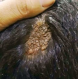
A sebaceous naevus is a congenital skin lesion which usually manifests as a bald patch on the scalp of the baby. The area of hair loss looks orange or yellowish, with a velvety feel and appearance. It is usually oval or circular. In adolescence, under the influence of sex hormones, it becomes bumpy and warty, with an unpleasant verrucoid appearance.
Throughout teenage years and early adulthood they usually stay quiescent. Sometimes they enlarge in later adulthood and, rarely, undergo neoplastic change which on the whole involves benign tumours.
Sebaceous naevi are hamartomas, meaning they arise from tissues normally found at that site but they grow in a disorganised way. They have a common embryological origin of the pilosebaceous-apocrine unit and therefore contain sebaceous glands, hair follicles and sweat glands. Hence the term sebaceous naevus is a little inaccurate: some pathologists prefer the term verrucous epidermal naevus or organoid naevus.1 Although they contain elements of hair follicles, normal terminal hair follicles are absent making the area look hairless. Most are solitary, with the scalp being the most common site.
Scalp sebaceous naevus
Sebaceous naevus (Epidemiology)
Estimated prevalence is 0.3% of all newborns worldwide, with an equal sex distribution.2
Sebaceous naevus symptoms (presentation)
Usually, a single hairless patch (round or linear) is noted on the scalp at birth, or shortly thereafter.
The classical velvety tan or yellow-orange appearance may be noted.
The lesion may appear more prominent in newborns, due to the effects of maternal sex hormones.
In adolescents the lesion tends to take on its nodular verrucoid appearance due to the influence of sex hormones.
In later life there may be overgrowth of components of the lesion indicating possible benign or malignant tumours developing within the lesion.
Differential diagnosis
The appearance of sebaceous naevi is fairly characteristic but the following lesions may have a similar appearance or affect similar areas of the body:
Aplasia cutis congenita.
Naevus syringocystadenomatosus papilliferous.
Juvenile xanthogranulomata.
Solitary mastocytoma.
Other causes of alopecia.
Investigations
Biopsy may be considered for diagnostic purposes or to investigate the nature of tumours that arise within them.
Associated diseases
About a third of people with a sebaceous naevus have systemic abnormalities affecting the brain, eye or skeletal systems. When these occur it is termed sebaceous naevus syndrome.3 In these cases the naevi follow the lines of Blaschko; the more extensive the cutaneous involvement, the greater the risk of extracutaneous manifestations.4 Subsequent problems can include infantile epilepsy, developmental delay and intellectual disability.
Sebaceous naevus treatment and management5
Due to the difficulty of quantifying the risk of malignant transformation, some specialists advocate full-thickness skin excision before adolescence.6 Others recommend watchful waiting.5
Some cases will in any case not be suitable for excision due to their size and location. These should be subjected to regular review.
Photodynamic therapy can play a role in removing the lesion.7
Seek the advice of a dermatologist and/or plastic surgeon to help decide on the need for, and timing of, excision.
Any lesion that changes its appearance significantly, ulcerates, becomes painful, bleeds or shows other signs of potential malignancy, should be referred to a dermatologist.
Complications
Poor cosmetic appearance.
Development of a benign or malignant tumour within the lesion.
Complications associated with removal of the lesion.
Prognosis
Most lesions remain quiescent throughout adult life, the only concern being cosmetic. Two case series studying nearly 1,300 sebaceous naevi have found that 15% undergo neoplastic change.8 9
Of these 15%, 80% develop foci of benign neoplasms such as trichoblastomas and syringocystadenomas. The remaining 20% develop malignant changes, with basal cell carcinomas making up half of those, squamous cell carcinomas a quarter.
Usually, any malignant change occurs in adulthood but it has been reported in children as young as 8 years.6 Malignancy can be diagnosed in the same way as other malignant skin tumours, with dermoscopy playing its usual role.10
Patient picks for Dermatology
Professional
Epidermal naevus and its syndromes
Synonyms: Jadassohn's naevus phakomatosis Background Epidermal naevi; Primary Care Dermatology Society. September 2022. 68153 : PCDS Epidermal naevi Epidermal Nevus; D... Naevus sebaceous Sebaceous naevi are relatively common among ENS. Secondary tumours may occur in about 25% of naevus sebaceous, most of which are benign (eg, trichoblastomas,.
by Dr Colin Tidy, MRCGP

Professional
Spider naevus
Synonyms: arterial spider, vascular spider, naevus araneus, naevus arachnoidius, spider angioma, tache stellaire, étoile vasculaire. What is spider naevus. Spider naevi is the name given to small angiomata which appear on the surface of the skin. They were first described in 1869 by the English physician Erasmus Wilson. Hadley, R; The Life and.
by Dr Colin Tidy, MRCGP
Professional
Pilar cysts
What are epidermoid and pilar cysts. Epidermoid or pilar cysts are very common and are usually of no clinical significance. Epidermoid cysts (also known as epithelial, epidermal or sebaceous cysts) are intradermal or subcutaneous tumours. Epidermoid cysts may occur anywhere on the body but occur most often on the face, scalp, neck, back and.
by Dr Philippa Vincent, MRCGP
Professional
Congenital pigmented naevus
Synonyms: congenital melanocytic naevus, naevocellular naevus, giant hairy naevus A congenital melanocytic naevus is a proliferation of benign melanocytes that are present at birth or develop shortly after birth. Congenital melanocytic naevi, DermNet NZ 21769 : DermNet NZ Congenital melanocytic naevi Congenital pigmented naevi initially appear as.
by Dr Colin Tidy, MRCGP

Professional
Becker's naevus
Synonyms: Becker melanosis, pigmented hairy naevus of Becker Becker's naevus is a form of epidermal naevus (birthmark). Becker Naevus; DermNet. Becker's naevus syndrome is a type of epidermal naevus syndrome. It is characterised by the presence of a Becker's naevus with ipsilateral breast hypoplasia or hypoplastic defects of the muscle, skin, or.
by Dr Colin Tidy, MRCGP

Professional
Strawberry naevus
Synonym: infantile haemangioma What is strawberry naevus. Strawberry naevus is the name given to an haemangioma which occurs in infancy, usually on the face. Natural history Infantile haemangiomas (including strawberry naevi); Primary Care Dermatology Society. 62593 : PCDS Infantile haemangiomas (including strawberry naevi) The strawberry naevus.
by Dr Colin Tidy, MRCGP
Exclusive updates for healthcare professionals
Stay informed with the latest clinical updates, professional insights, and evidence-based guidance. The Patient Pro newsletter curates essential content for healthcare professionals—delivered straight to your inbox.
By subscribing you accept our Privacy Policy. You can unsubscribe at any time. We never sell your data.
Further reading and references
- Simi CM, Rajalakshmi T, Correa M; Clinicopathologic analysis of 21 cases of nevus sebaceus: a retrospective study. Indian J Dermatol Venereol Leprol. 2008 Nov-Dec;74(6):625-7.
- Moody MN, Landau JM, Goldberg LH; Nevus sebaceous revisited. Pediatr Dermatol. 2012 Jan-Feb;29(1):15-23. doi: 10.1111/j.1525-1470.2011.01562.x. Epub 2011 Oct 13.
- Laura FS; Epidermal nevus syndrome. Handb Clin Neurol. 2013;111:349-68. doi: 10.1016/B978-0-444-52891-9.00041-5.
- Asch S, Sugarman JL; Epidermal nevus syndromes. Handb Clin Neurol. 2015;132:291-316. doi: 10.1016/B978-0-444-62702-5.00022-6.
- Sebaceous naevus; DermNet NZ
- Rosen H, Schmidt B, Lam HP, et al; Management of nevus sebaceous and the risk of Basal cell carcinoma: an 18-year review. Pediatr Dermatol. 2009 Nov-Dec;26(6):676-81. doi: 10.1111/j.1525-1470.2009.00939.x. Epub 2009 Jul 20.
- Babilas P, Szeimies RM; The use of photodynamic therapy in dermatology. G Ital Dermatol Venereol. 2010 Oct;145(5):613-30.
- Idriss MH, Elston DM; Secondary neoplasms associated with nevus sebaceus of Jadassohn: a study of 707 cases. J Am Acad Dermatol. 2014 Feb;70(2):332-7. doi: 10.1016/j.jaad.2013.10.004. Epub 2013 Nov 20.
- Cribier B, Scrivener Y, Grosshans E; Tumors arising in nevus sebaceus: A study of 596 cases. J Am Acad Dermatol. 2000 Feb;42(2 Pt 1):263-8.
- Enei ML, Paschoal FM, Valdes G, et al; Basal cell carcinoma appearing in a facial nevus sebaceous of Jadassohn: dermoscopic features. An Bras Dermatol. 2012 Jul-Aug;87(4):640-2.
About the authorView full bio
Updated by

Dr Doug McKechnie, MRCGP
Medical Writer
MA, MBBS, MSc, DRCOG, MRCP(UK), MRCGP(2021), FHEA
Dr Doug McKechnie is an NHS GP working in London. He works full-time clinically and is also the Deputy Lead for the Clinical and Professional Practice module at University College London Medical School.
About the reviewerView full bio

Dr Philippa Vincent, MRCGP
General Practitioner, Medical Author
MB BS, Bsc, MRCGP (2000), DCH, DFSRH, DRCOG
Dr Philippa Vincent is an NHS GP working in North London.
Simple access to trusted treatments and health services online.
Common conditions
Get help with common conditions through the NHS, without a GP appointment.





More in dermatology
- Becker's naevus
- Chronic plaque psoriasis
- Cicatricial pemphigoid
- Cold injury
- Dermatophytosis
- Diabetic foot
- Epidermolysis bullosa
- Erythema annulare centrifugum
- Erythema induratum
- Febrile neutrophilic dermatosis
- Folliculitis
- Hidradenitis suppurativa
- Lichen simplex
- Pathogenic free-living amoeba
- Rubella
- Smallpox
- Stevens-Johnson syndrome
- Sunburn
- Systemic sclerosis
- Vitiligo