Ankylosing spondylitis
Peer reviewed by Dr Doug McKechnie, MRCGPLast updated by Dr Philippa Vincent, MRCGPLast updated 14 Mar 2024
Meets Patient’s editorial guidelines
- DownloadDownload
- Share
- Language
- Discussion
- Audio Version
- Add to preferred sources on Google
Medical Professionals
Professional Reference articles are designed for health professionals to use. They are written by UK doctors and based on research evidence, UK and European Guidelines. You may find the Ankylosing spondylitis article more useful, or one of our other health articles.
What is ankylosing spondylitis?
Synonyms: rheumatoid spondylitis, Marie-Strümpell disease, von Bechterew's disease
Ankylosing spondylitis (AS) is a chronic seronegative spondyloarthropathy which primarily involves the axial skeleton (ie sacroiliitis and spondylitis). The aetiology is unknown but involves the interaction of genetic and environmental factors. The diagnosis is made by combining clinical criteria of inflammatory back pain and enthesitis (inflammation at the site of bone insertion of ligaments and tendons) or arthritis with radiological findings.
It is thought that ankylosing spondylitis is triggered by an environmental factor (or factors) in people who are genetically predisposed.1
Axial spondyloarthritis includes ankylosing spondylitis (radiographic axial spondyloarthritis) and non-radiographic axial spondyloarthritis. See also the separate Axial spondyloarthritis article for further details, including a summary of the current National Institute for Health and Care Excellence (NICE) clinical guideline.2
How common is ankylosing spondylitis? (Epidemiology)13
The prevalence of AS is believed to range from 0.05% to 0.23%.
Estimates of the prevalence vary between countries, with mean prevalence per 10,000 of 31.9 in North America, 23.8 in Europe, 16.7 in Asia, 10.2 in Latin America and 7.4 in Africa.
Around twice as many men have AS compared with women.
Non-radiographic axial spondyloarthritis affects a similar number of women as men.
AS most commonly begins between 20 and 30 years of age, with 90-95% of people aged less than 45 years at disease onset.
80% of people develop the first symptoms before the age of 30 and less than 5% are diagnosed after the age of 45.
More than 90% heritability has been estimated for axial spondyloarthritis. The most important genetic risk factor is human leukocyte antigen B27 (HLA-B27). The prevalence of HLA-B27 usually reflects the prevalence of axial spondyloarthritis within a population. However axial spondyloarthritis can occur in people without HLA-B27.
Symptoms of ankylosing spondylitis (presentation)
Symptoms may be subtle in early stages or mild disease, with an insidious onset over several months to years.
AS usually presents before the age of 30 years.
Most patients have mild chronic disease or intermittent flares with periods of remission.
Systemic features are common. Fever and weight loss may occur during periods of active disease. Fatigue is also prominent.
Morning stiffness is characteristic.
Inflammatory back pain:
Often improves with moderate physical activity with no relief from rest.
Unlike mechanical back pain, patients often experience stiffness and pain which awaken them in the early morning hours.
The spinal disease starts in the sacroiliac joints (bilateral lumbosacral region) and may be felt as diffuse nonspecific buttock pain.
On examination there is often tenderness of the sacroiliac joints or a limited range of spinal motion.
In the advanced stages, patients develop loss of lumbar lordosis, buttock atrophy, and an exaggerated thoracic kyphosis with a stooped forward neck sometimes referred to as a 'question mark posture'.
Peripheral enthesitis:
Occurs in approximately a third of patients.
Common sites - behind the heel (Achilles tendonitis), the heel pad (plantar fasciitis) and the tibial tuberosity.
Lesions tend to be painful, especially in the morning. There may be associated swelling of the tendon or ligament insertion.
Peripheral arthritis:
Also occurs in about a third of patients.
Joint involvement is usually asymmetrical, involving the hips, shoulder girdle (glenohumeral, acromioclavicular and sternoclavicular joints), joints of the chest wall (costovertebral joints, costosternal junctions) and symphysis pubis.
Other peripheral joints are less often and less severely affected, usually as asymmetrical oligoarthritis.
In children, AS tends to commence with arthritis prior to spinal disease developing.
Temporomandibular joints are occasionally involved.
Examination
Measure chest expansion, lateral lumbar flexion and forward lumbar flexion.
Schober's test - see the separate article Examination of the spine, which deals with thoracolumbar back examination.
Palpate and stress the sacroiliac joints.
Examine peripheral joints for synovitis or enthesitis.
Always look for extra-articular manifestations of AS, as these occur in up to 40% of patients.
Extra-articular manifestations1 4
Extra-articular manifestations are common. A systematic review found a pooled prevalence for:
Uveitis of 25.8%. Occurs particularly in males and people who are HLA-B27 positive.
Psoriasis of 9.3%.
Inflammatory bowel disease (Crohn's disease or ulcerative colitis) of 6.8%.
Other extra-articular manifestations include:
Cardiac complications include:
Aortic regurgitation, mitral regurgitation and non-aortic valvular heart disease.
Arrhythmias may also be present (for example, ventricular and supraventricular extrasystoles).
Increased risk of myocardial infarction (and stroke).5 It is therefore important to manage modifiable cardiovascular risk factors where present (for example, smoking, raised body mass index (BMI), cholesterol, sedentary lifestyle, comorbidities).
Lung involvement such as restrictive pulmonary disease and apical fibrosis. Dyspnoea may occur as a result of costovertebral involvement decreasing vital capacity.
Neurological involvement from vertebral fracture, dislocation, or cauda equina syndrome (sensory disturbance in lower limbs and perineum).
Renal involvement: amyloidosis is a very rare complication in patients with severe, active and long-standing disease and may cause renal dysfunction with proteinuria and renal insufficiency or chronic kidney disease. Immunoglobulin A (IgA) nephropathy is another association.
Referral for suspected axial spondyloarthritis
NICE recommends referral if:2
Low back pain started before the age of 45 years and has lasted for longer than three months: refer the person to a rheumatologist for a spondyloarthritis assessment if four or more of the following additional criteria are also present:
Low back pain started before the age of 35 years (this further increases the likelihood that back pain is due to spondyloarthritis compared with low back pain that started between 35 and 44 years).
Waking during the second half of the night because of symptoms.
Buttock pain.
Improvement with movement.
Improvement within 48 hours of taking non-steroidal anti-inflammatory drugs (NSAIDs).
A first-degree relative has spondyloarthritis.
There is current or past arthritis.
There is current or past enthesitis.
There is current or past psoriasis.
If exactly three of the additional criteria are present, perform an HLA-B27 test. If the test is positive, refer the person to a rheumatologist for a spondyloarthritis assessment.
If the person does not meet these criteria but clinical suspicion of axial spondyloarthritis remains, advise the person to seek repeat assessment if new signs, symptoms or risk factors develop. This may be especially appropriate if the person has current or past inflammatory bowel disease (Crohn's disease or ulcerative colitis), psoriasis or uveitis.
Urgently refer people with suspected new-onset inflammatory arthritis to a rheumatologist for a spondyloarthritis assessment, unless rheumatoid arthritis, gout or acute calcium pyrophosphate arthritis ('pseudogout') is suspected. If rheumatoid arthritis is suspected, see referral for specialist treatment in the NICE guideline on rheumatoid arthritis in adults.
Refer people with dactylitis to a rheumatologist for a spondyloarthritis assessment.
Refer people with enthesitis without apparent mechanical cause to a rheumatologist for a spondyloarthritis assessment if:
It is persistent.
It is in multiple sites.
Any of the following are also present:
Back pain without apparent mechanical cause.
Current or past uveitis. Refer people for an immediate (same-day) ophthalmological assessment if they have symptoms of acute anterior uveitis (for example, eye pain, eye redness, sensitivity to light or blurred vision).
Current or past psoriasis.
Gastrointestinal or genitourinary infection.
Inflammatory bowel disease.
A first-degree relative has spondyloarthritis or psoriasis.
Diagnosis1
Modified New York criteria for diagnosing ankylosing spondylitis:
Clinical criteria:
Low back pain; present for more than three months; improved by exercise but not relieved by rest.
Limitation of lumbar spine motion in both the sagittal and frontal planes.
Limitation of chest expansion relative to normal values for age and sex.
Radiological criterion: sacroiliitis on X-ray.
Diagnose: definite AS if the radiological criterion is present plus at least one clinical criterion. Probable AS if three clinical criteria are present alone, or if the radiological criterion is present but no clinical criteria are present.
Associated diseases
AS may overlap with other spondyloarthropathies - for example, psoriatic arthritis, reactive arthritis or enteropathic arthropathy.
Differential diagnosis1
Inflammatory conditions - for example, rheumatoid arthritis, psoriatic arthritis, reactive arthritis.
Degenerative conditions - for example, osteoarthritis, degenerative disc disease, spondylosis, degenerative changes in the intervertebral (facet) joints.
Infection - for example, tuberculosis.
Neoplasms, primary or secondary.
Congenital spinal deformity.
Fractures.
Infectious sacroiliitis.
Spinal stenosis.
Referred pain.
Diagnosing ankylosing spondylitis (investigations)
Blood tests
No laboratory tests are specific and are often more helpful to exclude other diagnoses rather than confirming AS.
However, 50-70% of people with AS will have raised CRP and ESR.3
Referral to a rheumatologist should be made if the patient has 4 or more diagnostic criteria or the patient has 3 diagnostic criteria plus is HLA-B27 positive.1
Imaging
X-rays are the most helpful imaging modality in established disease, although they may be normal in early disease.
Look for sacroiliitis or enthesitis (particularly of the annulus fibrosus). Sacroiliitis initially shows as blurring in the lower part of the joint, then bony erosions or sclerosis occur and widening or eventual fusion of the joint.
The vertebral bodies may become 'squared'. In later stages, bony bridges (syndesmophytes) form between adjacent vertebrae, there is ossification of spinal ligaments and, in late disease, there may be complete fusion of the vertebral column (bamboo spine).
Spinal osteopenia is common.
MRI scanning may be useful in identifying early sacroiliitis. MRI of the sacroiliac joints is more sensitive than either plain X-ray or CT scan in demonstrating sacroiliitis. It has a growing role in diagnosis, prognostication and selection of patients for biological treatment but is not recommended for primary care.
MRI/CT scans - useful in making the diagnosis of a spinal fracture in patients with late-stage spinal disease.
Dual-energy X-ray absorptiometry (DXA) scans are used to assess for osteoporosis but may underestimate the fracture risk in AS, due to new bone formation in the spine.
Musculoskeletal ultrasound scanning can help in diagnosing enthesitis.6
Bamboo spine
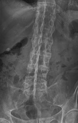
© By Stevenfruitsmaak, via Wikimedia Commons
Management of ankylosing spondylitis1 7
General
Ankylosing spondylitis is a chronic condition for which there is currently no cure. There are wide individual differences in the impact of AS and the aim of treatment is essentially symptomatic with good control of symptoms, maintenance of function (facilitated by early diagnosis) and management of complications.
Refer all new or suspected cases of AS to a rheumatologist. This is for confirmation of the diagnosis, review of current treatment, access to specialist physiotherapy and occupational therapy and the arrangement of follow-up (often as part of a shared care arrangement with primary care).
Treatment of extra-articular manifestations may require the involvement of other specialist teams.
Physiotherapy and rehabilitation
Physiotherapy, including an exercise programme and postural training, is important to maintain function and, in some severe cases, a period of inpatient intensive rehabilitation may be warranted.
A Cochrane review found that an individual home-based or supervised exercise programme is better than no intervention; that supervised group physiotherapy is better than home exercises; and that combined inpatient spa-exercise therapy followed by group physiotherapy are better than group physiotherapy alone.8
Spinal extension and deep-breathing exercises help to maintain spinal mobility, encourage erect posture and promote chest expansion.
Maintaining an erect posture during daily activities and sleeping on a firm mattress with a thin pillow also tend to reduce the tendency towards thoracic kyphosis.
Hydrotherapy and swimming are excellent activities to maintain mobility and fitness.
Anti-inflammatories9
Non-steroidal anti-inflammatory drugs (NSAIDs) improve the symptoms of the disease.10 Commence treatment with an NSAID unless contra-indicated and, in those at increased risk of gastrointestinal side-effects, consider the combination of an NSAID and proton pump inhibitor (PPI) or cyclo-oxygenase-2 (COX-2) inhibitor and PPI. Where contra-indicated or poorly tolerated, a standard analgesic such as paracetamol +/- codeine should be substituted.1
Where NSAIDs do not control symptoms sufficiently:
Consider a slow-release preparation if morning stiffness is a particular issue.
Add additional pain relief (for example, simple analgesics, amitriptyline) where there is poor sleep due to pain.
Local corticosteroid injections are useful for symptomatic sacroiliitis, peripheral enthesitis and arthritis.
Oral corticosteroids are not recommended in ankylosing spondylitis.3
Refer to a rheumatologist for consideration of additional therapy.
Cytokine modulators11
TNF-alpha inhibitors are effective in AS. They should only be used under the care of a rheumatologist.
NICE guidance:12
Adalimumab, certolizumab pegol, etanercept, golimumab and infliximab are options for treating severe active AS in adults whose disease has responded inadequately to, or who cannot tolerate, non-steroidal anti-inflammatory drugs.
The response to adalimumab, certolizumab pegol, etanercept, golimumab or infliximab treatment should be assessed 12 weeks after the start of treatment. Treatment should only be continued if there is clear evidence of response.
Treatment with another tumour necrosis factor (TNF) alpha inhibitor is recommended for people who cannot tolerate, or whose disease has not responded to, treatment with the first TNF-alpha inhibitor, or whose disease has stopped responding after an initial response.
The use of TNF-alpha inhibitors is known to increase the risk of serious infections.13
NICE recommends the use of secukinumab, ixekizumab and upadacitinib as options for treating active AS in adults whose disease has responded inadequately to conventional therapy (NSAIDs or TNF-alpha inhibitors). Response should be assessed after 16 weeks of treatment and only continued if there is clear evidence of response.14 15 16
Surgery17
Untreated AS can cause spinal deformity, with more than 30% of AS patients suffering from thoracolumbar kyphosis. Corrective osteotomy and stabilisation are recommended under certain conditions, such as severe kyphosis or advanced hip arthritis.
Complications of ankylosing spondylitis
The complications of axial spondyloarthritis include: 1317
Ankylosis or spinal fusion resulting from new bone formation.
Spinal fractures.
Hip involvement, affecting about a third of people with AS and may require joint replacement.
Adverse effects from treatment - for example, NSAIDs (gastritis, ulcers, renal effects), biological DMARDs (infection, immunosuppression, malignancy).
Mood disorders.
Decreased quality of life and work productivity due to pain, stiffness, fatigue, reduction in spinal mobility and physical function, and sleep problems.
Prognosis1
The course is variable but damage is progressive and irreversible.
The prognosis also depends on the presence of extraspinal manifestations (for example, uveitis, psoriasis, inflammatory bowel disease), age at diagnosis, and the choice of treatment.
Increased risk of spinal fracture later in life. The spine is made more brittle by rigidity and weaker by osteoporosis. Resultant fractures can occur with minimal force.
Exclusive updates for healthcare professionals
Stay informed with the latest clinical updates, professional insights, and evidence-based guidance. The Patient Pro newsletter curates essential content for healthcare professionals—delivered straight to your inbox.
By subscribing you accept our Privacy Policy. You can unsubscribe at any time. We never sell your data.
Further reading and references
- Axial spondyloarthritis (including ankylosing spondylitis); NICE CKS, April 2024 (UK access only)
- Spondyloarthritis in over 16s: diagnosis and management; NICE Guidance (Feb 2017)
- Wenker KJ, Quint JM; Ankylosing Spondylitis.
- El Maghraoui A; Extra-articular manifestations of ankylosing spondylitis: prevalence, characteristics and therapeutic implications. Eur J Intern Med. 2011 Dec;22(6):554-60. Epub 2011 Jul 13.
- Mathieu S, Pereira B, Soubrier M; Cardiovascular events in ankylosing spondylitis: An updated meta-analysis. Semin Arthritis Rheum. 2014 Oct 18. pii: S0049-0172(14)00248-0. doi: 10.1016/j.semarthrit.2014.10.007.
- Hamdi W, Chelli-Bouaziz M, Ahmed MS, et al; Correlations among clinical, radiographic, and sonographic scores for enthesitis in ankylosing spondylitis. Joint Bone Spine. 2011 May;78(3):270-4. Epub 2010 Oct 30.
- van der Heijde D, Ramiro S, Landewe R, et al; 2016 update of the ASAS-EULAR management recommendations for axial spondyloarthritis. Ann Rheum Dis. 2017 Jun;76(6):978-991. doi: 10.1136/annrheumdis-2016-210770. Epub 2017 Jan 13.
- Dagfinrud H, Kvien TK, Hagen KB; Physiotherapy interventions for ankylosing spondylitis. Cochrane Database Syst Rev. 2008 Jan 23;(1):CD002822.
- Sidiropoulos PI, Hatemi G, Song IH, et al; Evidence-based recommendations for the management of ankylosing spondylitis: systematic literature search of the 3E Initiative in Rheumatology involving a broad panel of experts and practising rheumatologists. Rheumatology (Oxford). 2008 Mar;47(3):355-61.
- Kroon FP, van der Burg LR, Ramiro S, et al; Nonsteroidal Antiinflammatory Drugs for Axial Spondyloarthritis: A Cochrane Review. J Rheumatol. 2016 Mar;43(3):607-17. doi: 10.3899/jrheum.150721. Epub 2016 Feb 1.
- Updates on ankylosing spondylitis: pathogenesis and therapeutic agents; Journal of Rheumatic Diseases
- TNF-alpha inhibitors for ankylosing spondylitis and non-radiographic axial spondyloarthritis; NICE Technology Appraisal Guidance, February 2016
- Minozzi S, Bonovas S, Lytras T, et al; Risk of infections using anti-TNF agents in rheumatoid arthritis, psoriatic arthritis, and ankylosing spondylitis: a systematic review and meta-analysis. Expert Opin Drug Saf. 2016 Dec;15(sup1):11-34. doi: 10.1080/14740338.2016.1240783.
- Secukinumab for active ankylosing spondylitis after treatment with non-steroidal anti-inflammatory drugs or TNF-alpha inhibitors; NICE Technology appraisal guidance, September 2016
- Ixekizumab for treating axial spondyloarthritis; NICE Technology appraisal guidance, July 2021
- Upadacitinib for treating active ankylosing spondylitis; NICE Technology appraisal guidance, September 2022
- Zhu W, He X, Cheng K, et al; Ankylosing spondylitis: etiology, pathogenesis, and treatments. Bone Res. 2019 Aug 5;7:22. doi: 10.1038/s41413-019-0057-8. eCollection 2019.
About the authorView full bio

Dr Philippa Vincent, MRCGP
General Practitioner, Medical Author
MB BS, Bsc, MRCGP (2000), DCH, DFSRH, DRCOG
Dr Philippa Vincent is an NHS GP working in North London.
About the reviewerView full bio

Dr Doug McKechnie, MRCGP
Medical Writer
MA, MBBS, MSc, DRCOG, MRCP(UK), MRCGP(2021), FHEA
Dr Doug McKechnie is an NHS GP working in London. He works full-time clinically and is also the Deputy Lead for the Clinical and Professional Practice module at University College London Medical School.
Article history
The information on this page is written and peer reviewed by qualified clinicians.
Article also available in English, German, Spanish, French, Italian, Portuguese, Hindi, Hebrew, Arabic, and Swedish.
Next review due: 13 Mar 2028
14 Mar 2024 | Latest version

Ask, share, connect.
Browse discussions, ask questions, and share experiences across hundreds of health topics.

Feeling unwell?
Assess your symptoms online for free
More in orthopaedics and sports medicine
- Acromioclavicular joint problems
- Ankle fractures
- Brachial plexus assessment and common injuries
- Brown-Séquard syndrome
- Classifying open fractures
- Elbow injuries and fractures
- Fat embolism syndrome
- Femoral fractures
- Gangrene
- Hip dislocations
- Kienböck's disease
- Klippel-Feil syndrome
- Musculocutaneous nerve lesion
- Osteoporosis
- Overuse phenomena and RSI
- Reactive arthritis
- Sever's disease
- Sports injuries - basic principles
- Tarsal tunnel syndrome
- Wrist fractures