Epidermal naevus and its syndromes
Peer reviewed by Dr Toni Hazell, MRCGPLast updated by Dr Colin Tidy, MRCGPLast updated 22 Sept 2023
Meets Patient’s editorial guidelines
- DownloadDownload
- Share
- Language
- Discussion
Medical Professionals
Professional Reference articles are designed for health professionals to use. They are written by UK doctors and based on research evidence, UK and European Guidelines. You may find one of our health articles more useful.
In this article:
Synonyms: Jadassohn's naevus phakomatosis
Continue reading below
What is an epidermal naevus?1 2
A naevus is a non-neoplastic skin or mucosal lesion. It is an abnormal mixture of a tissue’s normal components, usually presenting at birth or at a young age. An epidermal naevus is a naevus predominantly composed of keratinocytes.
Epidermal naevi may present in a variety of forms - Becker's naevus, verrucous epidermal naevus, inflammatory linear verrucous epidermal naevus, naevus comedonicus, eccrine naevus, apocrine naevus and white sponge naevus.
The majority of epidermal naevi only affect the skin. However, rarely, they can be associated with a wide range of non-cutaneous abnormalities, especially the central nervous system, eyes and skeleton (epidermal naevus syndrome). The epidermal naevus syndromes usually arise sporadically, with the exception of CHILD syndrome, which is familial.3
Epidermal naevi are classified according to clinical morphology, site and extent of involvement as well as the predominating epidermal structure within the individual lesion. If there are extensive epidermal naevi then they can be associated with extradermal abnormalities as part of the epidermal naevus syndrome (ENS). These are a heterogeneous group of disorders characterised by the presence of:4
One or more congenital hamartomatous ectodermal naevi of the skin.
Other organ involvement - commonly brain or skeleton.5
The terminology used to describe ENS can vary from author to author. The following can be described on their phenotype.4 6 7
Naevus sebaceous
Sebaceous naevi are relatively common among ENS.
Skin lesions are salmon or yellow in colour and waxy.
Epilepsy and intellectual impairment may be associated.
Secondary tumours may occur in about 25% of naevus sebaceous, most of which are benign (eg, trichoblastomas, syringocystadenoma papilliferum or other basaloid proliferations), although malignant tumours can occur.8
Keratinocytic epidermal naevus
Again, relatively common.
Pink or hyperpigmented plaques.
Intellectual impairment, structural brain abnormalities, skeletal abnormalities and strabismus are reported.
Naevus comedonicus
Skin lesions are localised collections of dilated follicles containing keratin, which may become infected or inflamed.
Electroencephalogram (EEG) abnormalities, cataracts and skeletal abnormalities are reported.
Congenital hemidysplasia, ichthyosiform naevus and limb defects syndrome9
Synonym: congenital unilateral ichthyosiform erythroderma
CHILD = Congenital Hemidysplasia, Ichthyosiform Naevus and Limb Defects.
Skin lesions are unilaterally inflammatory, reddened patches which may have dry scales; they are often located in the skinfolds.
Ipsilateral hypoplasia of limbs can occur; the axial skeleton may also be affected.
EEG abnormalities and hemispheric brain hypoplasia and cardiac abnormalities are reported.
Becker's naevus (pigmented hairy epidermal naevus)
Becker's naevus is a patch of hyperpigmentation and hypertrichosis, which is androgen-dependent and so becomes more prominent after puberty in males; it is usually located on the shoulder.
Onset is usually in late childhood/adolescence.
It is associated with skeletal abnormalities, underlying muscle hypoplasia and ipsilateral hypoplasia of the breast.
Proteus syndrome
Variable clinical features, characterised by overgrowth of multiple tissues.
CNS involvement is not typical but seizures and intellectual impairment have been reported.
Other possible features are ovarian or parotid tumours, lipomas and vascular malformations.
There are diagnostic criteria.
Phacomatosis pigmentokeratotica
A speckled lentiginous naevus associated with an epidermal naevus. The melanocytic component usually follows a segmental pattern ('checkerboard pattern') rather than following the lines of Blaschko.
There may be neurological, ocular and skeletal abnormalities.
Hemiatrophy with muscle weakness is common.
Inflammatory linear verrucous epidermal naevus
Skin lesions are linear, pruritic, reddened and hyperkeratotic papules or plaques.
Usually unilateral on the lower half of the body, often the buttock.
Often presents at <6 months of age.
May resemble psoriasis.
There may be associated skeletal abnormalities, although this point has been questioned.
Porokeratotic eccrine naevus
Verrucous, keratotic papules on the palms and soles.
There may be widespread cutaneous involvement.
Epidermal naevus aetiology
The lesions probably arise from abnormal ectodermal cells in the embryo.10 Ectodermal cells give rise to the skin epidermis and to neural tissue. Mosaicism seems to be involved, ie there are genetically different cell lines in one individual, and the abnormal cells give rise to the lesions.
A detailed review of the genetic basis and pathogenesis of ENS is available.4
Continue reading below
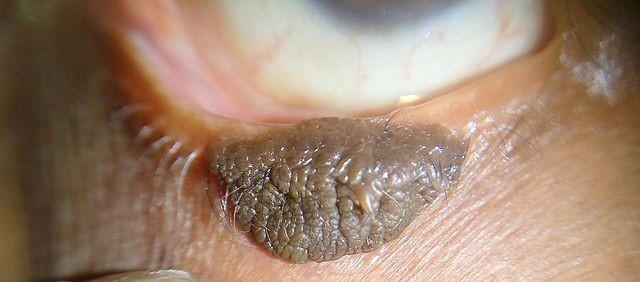
Visual appearance
Epidermal naevus

Imrankabirhossain, CC BY-SA 4.0, via Wikimedia Commons
Epidermal naevus epidemiology
Epidermal naevi (with or without other organ involvement) have an incidence of around 2 per 1,000 live births.4
Epidermal naevus syndromes are all very uncommon, without reliable population incidence figures. Of patients with epidermal naevi, 10-30% may have disorders in other organs suggesting an ENS.11 12
Continue reading below
Epidermal naevus symptoms
(Presentation)10
Epidermal naevi are present at birth (50%) or develop during childhood (mostly in the first year of life). They tend to grow during childhood and then stabilise during the teenage years.
Apart from sebaceous naevi (affecting the scalp), an epidermal naevus usually arises on the trunk and limbs and is uncommon on the face or scalp. The majority are linear epidermal naevi, which form a line, usually unilateral, which is also known as naevus unis lateralis. When they first appear, they are flat tan or brown marks, but as the child ages, they become thickened and often warty. The naevus may also become more extensive for a few years.
Systematised epidermal naevi are less common and are sometimes known as ichthyosis hystrix. There are multiple lesions that usually arise in a swirled pattern on one or both sides of the body. These may be associated with other congenital abnormalities (ENS).
The lesions themselves are usually asymptomatic apart from their appearance. However, inflammatory linear verrucous epidermal naevus lesions may be inflamed and irritated; naevus comedonicus lesions may become infected or inflamed.
In ENS, neurological involvement may include:
Epilepsy or infantile spasms.
Intellectual impairment.
Structural or vascular brain abnormalities.
Spinal lesions.
Skeletal involvement includes:
Incomplete formation of bony structures - eg, spina bifida.
Hypoplasia of bones.
Bony cysts.
Asymmetry of the skull or spine.
Spontaneous fractures and rickets.
Ophthalmic involvement includes:
Colobomas.
Strabismus.
Ptosis.
Nystagmus.
Corneal opacities.
Retinal changes.
Various other ocular abnormalities which have been described.
Endocrine features have been reported:
Hypophosphataemic vitamin D-resistant rickets has occurred in a number of cases.13
Precocious puberty has been described in several cases.
Syndrome of inappropriate antidiuretic hormone (SIADH) has been reported in one case.
Differential diagnosis
Naevus syringocystadenomatosus papilliferus.
Juvenile xanthogranulomata.
Solitary mastocytoma.
Multiple melanocytic naevi.
Other skin and CNS lesions - eg:
Tuberous sclerosis.
McCune-Albright syndrome.
Assessment
Initial assessment, to include exclusion of an ENS:4
Full developmental and family history.
Careful examination of all skin lesions. Skin biopsy may be required.
Neurological examination.
Ophthalmic examination.
Assess skeleton, symmetry and gait.
Consider if further assessment is required:
Generally, children with small epidermal naevi and no other findings do not need further investigation, although skin biopsy should be considered.4
Large epidermal naevi, especially in the head and neck, may merit CNS assessment.
Epidermal naevi should be fully assessed if there is a suspected syndrome involving other organs.
Investigations
These should be based on the needs of the individual patient but may include:14
Skin lesions should be referred for dermatological assessment and/or biopsied to ascertain their nature.
The epidermal naevus syndromes are diagnosed clinically and investigations may include skeletal survey, chest X-ray, CT or MRI, and molecular testing.3
Epidermal naevus treatment and management
A multidisciplinary approach is necessary to optimise the management of ENS, for which there is no cure.3
Skin lesions
Depending on the location and size of the lesions, excision may be merited for cosmetic reasons; however, this may not be feasible if underlying structures are involved.
Other possible treatments are:
Laser treatment may be successful in some cases.
Topical vitamin D (calcipotriol) may be helpful in treating inflammatory linear verrucous epidermal naevus, although there is conflicting evidence about its effectiveness.
5-fluorouracil was used with good results in one case of a large linear epidermal naevus.
Tacrolimus and fluocinonide, in combination, were successfully used in one case of inflammatory linear verrucous epidermal naevus.15
Shave excision followed by phenol peeling was used with good outcome in one case of verrucous epidermal naevus.
If there is new growth of the skin lesion, excisional biopsy may be indicated to rule out malignancy.
CNS lesions
Anticonvulsants for epilepsy.
Neurosurgery has been used in a few selected cases to control epilepsy.16
Complications
Poor cosmetic appearance.
Inflammation of the lesion in certain forms (inflammatory linear verrucous epidermal naevus).
Benign or malignant tumours arising within the lesion.
Prognosis
The prognosis is very good. They have the potential to have benign appendageal tumours arise within them (often syringocystadenoma papilliferum), or to transform into malignant tumours such as basal cell carcinoma or squamous cell carcinoma.
The lifetime risk of malignant transformation in sebaceous naevi is thought to be less than 1%.17 Malignant transformation of the lesion is rare before adolescence/adulthood. Verrucous epidermal naevi seem to have a much lower chance of forming tumours.
Further reading and references
- Nicholson CL, Daveluy S; Epidermal Nevus Syndromes. StatPearls, Jan 2023.
- Epidermal naevi; Primary Care Dermatology Society. September 2022.
- Epidermal Nevus; DermIS (Dermatology Information System)
- Epidermal naevus syndromes; DermNet.
- Sugarman JL; Epidermal nevus syndromes. Semin Cutan Med Surg. 2007 Dec;26(4):221-30.
- Flores-Sarnat L, Sarnat HB; Phenotype/genotype correlations in epidermal nevus syndrome as a neurocristopathy. Handb Clin Neurol. 2015;132:9-25. doi: 10.1016/B978-0-444-62702-5.00002-0.
- Asch S, Sugarman JL; Epidermal nevus syndromes. Handb Clin Neurol. 2015;132:291-316. doi: 10.1016/B978-0-444-62702-5.00022-6.
- Happle R; The group of epidermal nevus syndromes Part I. Well defined phenotypes. J Am Acad Dermatol. 2010 Jul;63(1):1-22; quiz 23-4.
- Aslam A, Salam A, Griffiths CE, et al; Naevus sebaceus: a mosaic RASopathy. Clin Exp Dermatol. 2014 Jan;39(1):1-6. doi: 10.1111/ced.12209.
- Mi XB, Luo MX, Guo LL, et al; CHILD Syndrome: Case Report of a Chinese Patient and Literature Review of the NAD[P]H Steroid Dehydrogenase-Like Protein Gene Mutation. Pediatr Dermatol. 2015 Nov-Dec;32(6):e277-82. doi: 10.1111/pde.12701. Epub 2015 Oct 13.
- Epidermal naevi; DermNet NZ
- Vidaurri-de la Cruz H, Tamayo-Sanchez L, Duran-McKinster C, et al; Epidermal nevus syndromes: clinical findings in 35 patients. Pediatr Dermatol. 2004 Jul-Aug;21(4):432-9.
- Adams D, Athalye L, Schwimer C, et al; A profound case of linear epidermal nevus in a patient with epidermal nevus syndrome. J Dermatol Case Rep. 2011 Jun 6;5(2):30-3.
- Kishida ES, Muniz Silva MA, da Costa Pereira F, et al; Epidermal nevus syndrome associated with adnexal tumors, spitz nevus, and hypophosphatemic vitamin D-resistant rickets. Pediatr Dermatol. 2005 Jan-Feb;22(1):48-54.
- Lambert DA, Giannouli E, Schmidt BJ; Postpolio syndrome and anesthesia. Anesthesiology. 2005 Sep;103(3):638-44.
- Mutasim DF; Successful treatment of inflammatory linear verrucous epidermal nevus with tacrolimus and fluocinonide. J Cutan Med Surg. 2006 Jan-Feb;10(1):45-7.
- Loddenkemper T, Alexopoulos AV, Kotagal P, et al; Epilepsy surgery in epidermal nevus syndrome variant with hemimegalencephaly and intractable seizures. J Neurol. 2008 Nov;255(11):1829-31. Epub 2008 Nov 13.
- Aguayo R, Pallares J, Casanova JM, et al; Squamous cell carcinoma developing in Jadassohn's sebaceous nevus: case report and review of the literature. Dermatol Surg. 2010 Nov;36(11):1763-8. doi: 10.1111/j.1524-4725.2010.01746.x.
Continue reading below
Article history
The information on this page is written and peer reviewed by qualified clinicians.
Next review due: 20 Sept 2028
22 Sept 2023 | Latest version

Ask, share, connect.
Browse discussions, ask questions, and share experiences across hundreds of health topics.

Feeling unwell?
Assess your symptoms online for free