Echocardiogram
Peer reviewed by Dr Colin Tidy, MRCGPLast updated by Dr Toni Hazell, FRCGPLast updated 22 Nov 2022
Meets Patient’s editorial guidelines
- DownloadDownload
- Share
- Language
- Discussion
- Audio Version
- Add to preferred sources on Google
An echocardiogram is an ultrasound scan of the heart. It is sometimes just called an 'echo'. Ultrasound is a very high-frequency sound that you cannot hear but it can be emitted and detected by special machines. The scan can give accurate pictures of the heart muscle, the heart chambers and structures within the heart such as the valves.
At a glance
An echocardiogram uses ultrasound to create moving pictures of your heart.
It can show how well your heart and its valves are working.
The test is painless, takes 15-30 minutes, and requires no special preparation.
A probe is placed on your chest with gel to send and receive ultrasound waves.
Different types exist, including Doppler, stress, and transoesophageal echocardiograms.
Results are sent to the doctor who requested the test.
What does an echocardiogram show?
An echocardiogram can be carried out for many different reasons. It may be done to check how well your heart is working after major heart problems such as a heart attack, or to look at how well the valves are moving inside the heart. An echocardiogram can also help to see any fluid that may have collected around the heart and may be used to see if symptoms such as shortness of breath are caused by a cardiac cause such as heart failure.
How accurate is a echocardiogram?
Interpreting an echocardiogram is a skilled job. The sonographer will interpret the images and write a report, which will be sent to the requesting clinician (GP or consultant). In some cases a consultant may want to carry out an echocardiogram themselves, to get a first-hand look at the images.
How is an echocardiogram done?
The test is painless and takes about 15-30 minutes. You may have to turn on to your side during the test so that the operator can scan the heart from different angles.
You will need to undress to the waist and lie on the couch. A probe is placed on your chest (it is a bit like a very thick blunt pen). Also, a substance called a contrast agent (lubricating jelly) is put on your chest so the probe makes good contact with the skin. The probe is connected by a wire to the ultrasound machine and monitor. Pulses of ultrasound are sent from the probe through the skin towards your heart. The ultrasound waves then 'bounce back' (echo) from the heart and various structures in the heart.
Echocardiogram procedure
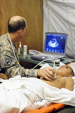
© Tech Sgt Luke Thelen, via Wikimedia Commons
The echoes are detected by the probe and are sent to the echocardiogram machine. They are displayed as a picture on the monitor. The picture is constantly updated so the scan can show movement as well as structure. (For example, the valves of a heart opening and closing.) The operator moves the probe around over the skin surface to obtain views from different angles. Some abnormalities can be seen quite clearly. For example, damaged heart valves, thickened heart muscle, some congenital heart defects, etc.
You do not need any special preparation before the test. You eat and drink normally before and after the test. Continue to take your usual medication.
Doppler echocardiography
This type of echocardiogram can measure variations in blood flow through your heart. For example, if can detect any abnormal blood flows next to a damaged valve. It can assess how well the heart valves are working. You do not need any special preparation before this test.
Stress echocardiogram
This test is done to show how well your heart responds to 'stress' such as exercise. In this test your doctor may do an echocardiogram, as described above, during or soon after exercise. Or you may be given a medication that causes the heart to beat harder and faster.
Transoesophageal echocardiogram
In this test you swallow a probe that is attached to a thin tube connecting it to an ultrasound machine. This views the heart from within the gullet (oesophagus) which lies just behind the heart. This can give a clearer view of the heart than normal echocardiography. It is done in situations where a very detailed picture is needed. For example, to assess valves before surgery is done to repair damaged valves, or to assess the extent of infection of a heart valve.
How long does it take to get echocardiogram results?
This varies locally. Results will go back to the requesting clinician and it is their responsibility to give them to the patient. So, if your echocardiogram was requested by a consultant, do not ring your GP for the result. Wait for your next consultant appointment, where you will get the result, or ring your consultant's secretary if you have a query. Hospital consultants should not ask patients to go to their GP for the results of tests that the consultant has requested.
Patient picks for Heart tests

Tests and investigations
Coronary angiography
Coronary angiography is a specialised X-ray test to find out detailed information about your heart (coronary) arteries. It is mainly used if you have angina or if you have had a heart attack, to assess which if any of the arteries are blocked, and how severely the arteries are blocked. It involves a procedure called catheterisation.
by Dr Doug McKechnie, MRCGP

Tests and investigations
Cardiac catheterisation
Cardiac catheterisation is a way to find out detailed information about your heart and coronary arteries and it is also possible to provide treatment for some conditions at the same time.
by Dr Colin Tidy, MRCGP
Frequently asked questions
What is the difference between a standard echocardiogram and a Doppler echocardiogram?
A standard echocardiogram primarily shows the structure and movement of your heart. A Doppler echocardiogram, however, specifically measures variations in blood flow through your heart. This allows it to detect abnormal blood flows, for example, next to a damaged valve, and assess how effectively the heart valves are working.
When would a transoesophageal echocardiogram be used instead of a standard one?
A transoesophageal echocardiogram is used when a very detailed picture of the heart is required. This is because the probe is swallowed and views the heart from inside your gullet, which is directly behind the heart, offering a clearer view than a standard echocardiogram where the probe is on the chest. It might be used to assess valves before surgery or to check the extent of a heart valve infection.
What happens after the echocardiogram test?
Following the test, the sonographer interprets the images and creates a report. This report is then sent to the clinician who requested the echocardiogram, such as your GP or a consultant. It is their responsibility to provide you with the results.
Do I need to do anything specific to prepare for an echocardiogram?
No, you do not need any special preparation for a standard, Doppler, or stress echocardiogram. You can eat and drink normally both before and after the test, and you should continue to take any regular medication as usual.
How long does an echocardiogram usually take?
The test itself is generally quick, typically taking around 15 to 30 minutes to complete.
Further reading and references
- Sengupta PP, Khandheria BK; Transoesophageal echocardiography. Heart. 2005 Apr;91(4):541-7.
- Mohamed AA, Arifi AA, Omran A; The basics of echocardiography. J Saudi Heart Assoc. 2010 Apr;22(2):71-6. doi: 10.1016/j.jsha.2010.02.011. Epub 2010 Mar 1.
About the authorView full bio

Dr Toni Hazell, FRCGP
MBBS, BSc, FRCGP, DFSRH, Dip GU med, DRCOG, DCH (London, UK, 2000)
Dr. Toni Hazell qualified from St. Mary’s Hospital Medical School and did her VTS at Northwick Park Hospital.
About the reviewerView full bio

Dr Colin Tidy, MRCGP
General Practitioner, Medical Author
MBBS, MRCGP, MRCP (Paediatrics), DCH
Dr Colin Tidy is an NHS Doctor, based in Oxfordshire.
Article history
The information on this page is written and peer reviewed by qualified clinicians.
Article also available in English, German, Spanish, French, Italian, Portuguese, Hindi, Hebrew, Arabic, and Swedish.
Next review due: 21 Nov 2027
22 Nov 2022 | Latest version

Ask, share, connect.
Browse discussions, ask questions, and share experiences across hundreds of health topics.

Feeling unwell?
Assess your symptoms online for free
Sign up to the Patient newsletter
Your weekly dose of clear, trustworthy health advice - written to help you feel informed, confident and in control.
By subscribing you accept our Privacy Policy. You can unsubscribe at any time. We never sell your data.
More in tests and investigations
- Amniocentesis
- Audiology
- Bowel cancer screening
- Breast screening
- Cardiac enzymes
- Cervical screening
- DMSA scan
- Exercise Tolerance Testing
- Faecal immunochemical test
- Gallium scan
- Home and ambulatory blood pressure recording
- Kidney biopsy
- Lumbar puncture
- MRCP scan
- Newborn bloodspot test
- Newborn physical examinations
- Osmolality, osmolarity and fluid homeostasis
- Sperm test
- Thyroid scans and uptake tests
- Ultrasound scan