Coronary angiography
Peer reviewed by Dr Colin Tidy, MRCGPLast updated by Dr Doug McKechnie, MRCGPLast updated 4 Mar 2025
Meets Patient’s editorial guidelines
- DownloadDownload
- Share
- Language
- Discussion
- Audio Version
- Add to preferred sources on Google
Coronary angiography is a specialised X-ray test to find out detailed information about your heart (coronary) arteries. It is mainly used if you have angina or if you have had a heart attack, to assess which if any of the arteries are blocked, and how severely the arteries are blocked. It involves a procedure called catheterisation.
At a glance
Coronary angiography is a procedure to look at the blood vessels that supply the heart.
During the procedure, a thin tube is inserted into an artery, and dye is injected to make the vessels visible on X-rays.
It can show the exact site and severity of any narrowing in the coronary arteries.
A coronary angioplasty, to treat narrowed arteries, can be done at the same time.
You will need to prepare for the procedure, which typically takes about 30 minutes.
Most side-effects are minor, but serious complications are rare.
What is a coronary angiography?
Coronary angiography is a medical procedure to look at the blood vessels that supply the heart (the coronary arteries). It is also called a coronary angiogram, cardiac catheterisation, or an invasive coronary angiogram.
Coronary angiography involves injecting dye into the coronary arteries. This dye makes the arteries visible on an X-ray. Dye is injected into the coronary arteries by using a catheter. (A catheter is a thin, flexible, hollow tube.) The catheter is inserted into a blood vessel in your arm or your groin, and passed up inside your blood vessels to reach the heart itself.
Coronary angiography can show the exact site and severity of any narrowing of the coronary arteries. This helps the doctor to decide on what treatment you may need. For example, if the narrowing is mild and does not need surgery; or, if the narrowing is severe and you should have a coronary artery bypass graft or coronary angioplasty (stent).
Coronary angioplasty (stenting) can be done at the same time as coronary angiography. They are often done together, if the angiography shows blood vessel narrowing that can be treated with angioplasty.
Computerised tomography (CT) coronary angiogram
A CT coronary angiogram is a different test to a traditional coronary angiogram.
A CT coronary angiogram uses a CT scanner to look at the heart and the coronary arteries. An injection of dye is given into a small vein in your hand or arm, and a CT scanner takes pictures of the heart and arteries.
A CT coronary angiogram doesn't require a catheter to be passed up to the heart, like a traditional coronary angiogram does. This means it is a less invasive test, and there are fewer risks compared to a traditional angiogram. However, it's not quite as good as a traditional angiogram at getting a very detailed picture of the coronary arteries. It's also not possible to perform treatments for a narrowed artery, such as angioplasty, at the same time.
A CT coronary angiogram is often used to rule out coronary heart disease. If the results are normal, this can avoid someone needing to have a traditional coronary angiogram. See separate leaflet called CT scan for more general information.
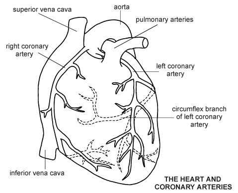
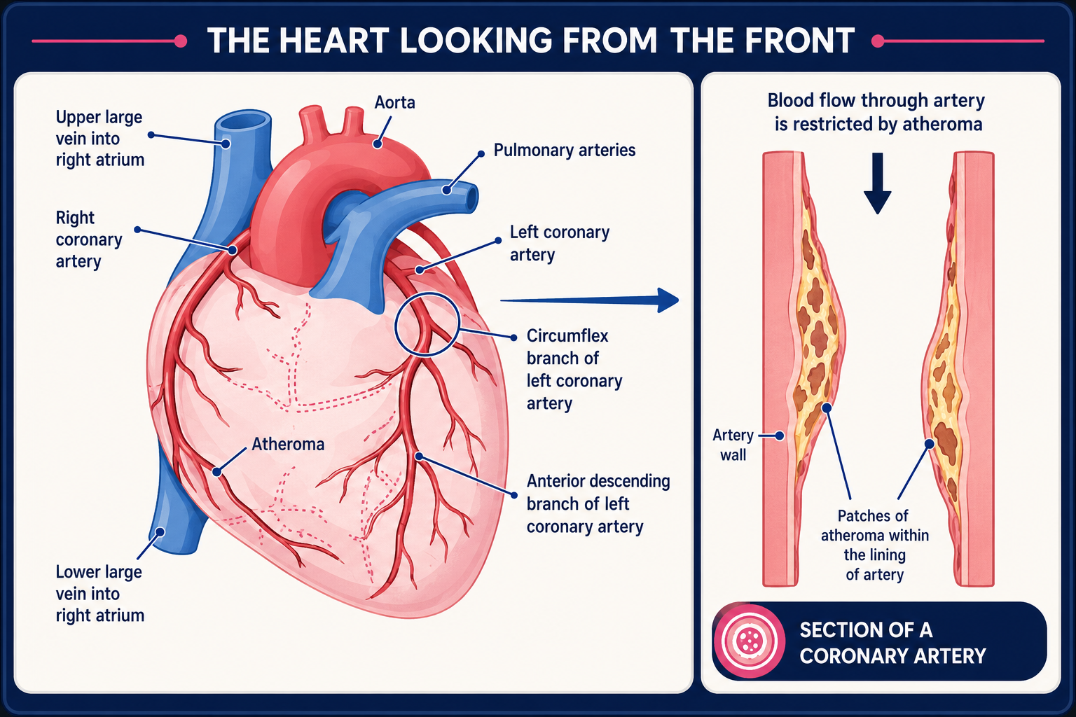
The heart and coronary arteries
Heart with atheroma
How is coronary angiography done?
You lie on a couch in a catheterisation room. An X-ray machine is mounted above the couch. A thin, flexible tube (a catheter) is inserted through a wide needle or small cut in the skin into a blood vessel in the groin or arm. Local anaesthetic is injected into the skin above the blood vessel. Therefore, it should not hurt when the catheter is passed into the blood vessel.
Coronary angiography
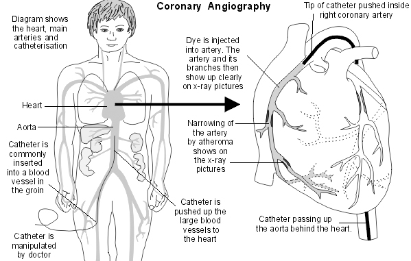
The doctor gently pushes the catheter up the blood vessel towards the heart. Low-dose X-rays are used to monitor the progress of the catheter tip which is gently manipulated into the correct position. You may be able to see the progress of the catheter on the X-ray monitor.
The tip of the catheter is pushed just inside a main coronary artery. Some dye is then injected down the catheter into the artery. Several X-ray films are rapidly taken as the dye is injected (the dye shows up clearly on X-ray films). The X-ray films are recorded as a moving picture and this is called an angiogram. The angiogram shows the vessels filling with blood and the sites of any narrowing can be seen.
The tip of the catheter is then put into the other main coronary artery and the test is repeated. So, an angiogram picture is built up of each of the coronary arteries and their branches.
You cannot feel the catheter inside the blood vessels. You may feel an occasional 'missed' or 'extra' heartbeat during the procedure. This is normal and doesn't cause any problems. During the procedure your heartbeat is monitored by electrodes placed on your chest which provide a tracing on an electrocardiograph (ECG) machine. Sometimes a sedative is given before the test if you are anxious.
If the angiogram has shown narrowing in the heart arteries, your cardiologist may proceed to open up the arteries (angioplasty) and possibly insert a stent as well, as part of the same procedure. See the coronary angioplasty leaflet for more details.
When the test is over, the catheter is gently pulled out. A dissolvable plug or a special stitch may be used to seal the puncture site. If it was inserted through a small cut in the skin in the arm then you will normally need a few stitches. Pressure might be used to stop the wound from bleeding, either by a nurse pressing on the site of insertion, or using a special pressure device.
How do I prepare for a coronary angiography?
You should receive instructions from your local hospital about what you need to do in the days leading up to the test. The sort of instructions may include:
Before the day of the test you may need a blood test and an ECG to make sure you are OK to have the procedure.
If you take a 'blood-thinning' medicine (anticoagulant), such as warfarin, you are likely to need to stop this for 2-3 days before the test. This prevents excessive bleeding from the site of the small, flexible tube (catheter) insertion.
If you take insulin or medicines for diabetes, the timing of when to take these on the day of the test may need to be clarified.
If you may be pregnant, you need to tell the doctor who will do the test.
You may be asked to stop eating and drinking for a few hours before the test.
You may be asked to shave both groins before the test.
You will have to sign a consent form at some point before the test to confirm that you understand the procedure, understand the possible complications -see below - and agree to the procedure being done.
How long does coronary angiography take?
It usually takes about 30 minutes. In most cases it is done as a day-case procedure.
After the test
The doctor will discuss what he or she found during the test. A letter is also sent to your doctor giving details of the test results.
You will need to rest for a few hours after the test. You should ask a friend or relative to accompany you home. Most people are able to resume their normal activities the next day.
There may be some bruising at the site of the small, flexible tube (catheter) insertion which may be a little sore when the anaesthetic wears off. Painkillers such as paracetamol will help to ease this.
You may need to have some stitches removed after about seven days if a small cut was made to insert the catheter.
Are there any risks or side-effects?
Most of the side-effects are minor and may include:
A bruise, which may form under the skin where the small, flexible tube (catheter) was inserted (usually the groin). This is not serious but it may be sore for a few days.
The small wound where the catheter is inserted sometimes becomes infected. Tell your doctor if the wound becomes red and tender. A short course of antibiotics will usually deal with this if it occurs.
Some people have a short angina-type pain during angiography. This soon goes.
The dye may give you a hot, flushing feeling when it is injected. Many people also describe a warm feeling in the groin when the dye is injected - as if they have 'wet themselves'. These feelings last just a few seconds (and the operator will tell you when they are about to inject the dye). Rarely, some people have an allergic reaction to the dye.
Complications
Serious complications are rare. Some people have a stroke or a heart attack (myocardial infarction) during the procedure. Also, rarely, the catheter may damage a heart (coronary) artery. The risk of serious complications is small and is mainly in people who already have serious heart disease. As a consequence of serious complications, some people have died during this procedure. Your doctor will only recommend coronary angiography if they feel the benefits outweigh the small risk.
Patient picks for Heart tests

Tests and investigations
Electrocardiogram
An electrocardiogram (ECG) records the electrical activity of the heart. The heart produces tiny electrical impulses which spread through the heart muscle to make the heart contract. These impulses can be detected by the ECG machine. An ECG may be used to help find the cause of symptoms such as the feeling of a 'thumping heart' (palpitations) or chest pain. Sometimes it is done as part of routine tests - for example, for high blood pressure, or before an operation. The ECG test is painless and harmless. (The ECG machine records electrical impulses coming from the body - it does not put any electricity into the body.)
by Dr Colin Tidy, MRCGP
Tests and investigations
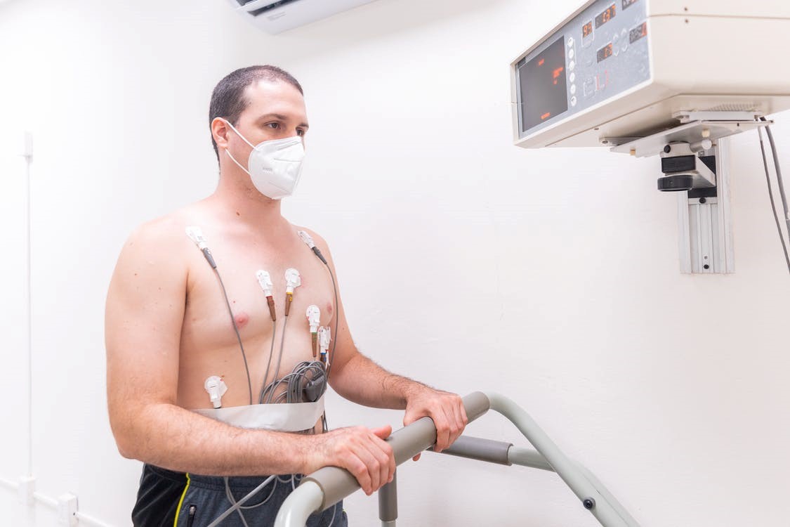
Exercise Tolerance Testing
An exercise tolerance test (ETT) records the electrical activity of the heart whilst exercising. It is most useful in patients who experience chest pain on exertion. It is also used to detect whether heart rhythm abnormalities can be brought on by exercise.
by Dr Philippa Vincent, MRCGP
Frequently asked questions
Will I be awake during a coronary angiogram?
Yes, you will typically be awake during the procedure. However, a sedative may be given if you are feeling anxious. Your heartbeat will be monitored throughout by electrodes placed on your chest.
What does a normal coronary angiogram result mean for a patient?
If a CT coronary angiogram shows normal results, it can help rule out coronary heart disease, potentially avoiding the need for a traditional coronary angiogram. For a traditional angiogram, if no significant narrowing is found, it means surgery or angioplasty may not be needed.
Can I eat or drink before a coronary angiogram?
You may be asked to stop eating and drinking for a few hours before the test. Specific instructions will be provided by your local hospital.
What should I do if I take blood-thinning medication?
If you take blood-thinning medicine (anticoagulant) like warfarin, you will likely need to stop it for 2-3 days before the test. This helps prevent excessive bleeding from the site where the catheter is inserted.
How soon can I return to normal activities after a coronary angiogram?
Most people are able to resume their normal activities the day after the procedure. You will need to rest for a few hours immediately after the test and should arrange for someone to accompany you home.
Further reading and references
- CT coronary angiography in patients with suspected angina due to coronary heart disease (SCOT-HEART): an open-label, parallel-group, multicentre trial; Lancet. 2015 Jun 13;385(9985):2383-91. doi: 10.1016/S0140-6736(15)60291-4. Epub 2015 Mar 15.
- Moss AJ, Newby DE; CT coronary angiographic evaluation of suspected anginal chest pain. Heart. 2015 Dec 8. pii: heartjnl-2015-307860. doi: 10.1136/heartjnl-2015-307860.
About the authorView full bio

Dr Doug McKechnie, MRCGP
Medical Writer
MA, MBBS, MSc, DRCOG, MRCP(UK), MRCGP(2021), FHEA
Dr Doug McKechnie is an NHS GP working in London. He works full-time clinically and is also the Deputy Lead for the Clinical and Professional Practice module at University College London Medical School.
About the reviewerView full bio

Dr Colin Tidy, MRCGP
General Practitioner, Medical Author
MBBS, MRCGP, MRCP (Paediatrics), DCH
Dr Colin Tidy is an NHS Doctor, based in Oxfordshire.
Article history
The information on this page is written and peer reviewed by qualified clinicians.
Article also available in English, German, Spanish, French, Italian, Portuguese, Hindi, Hebrew, Arabic, and Swedish.
Next review due: 3 Mar 2028
4 Mar 2025 | Latest version

Ask, share, connect.
Browse discussions, ask questions, and share experiences across hundreds of health topics.

Feeling unwell?
Assess your symptoms online for free
Sign up to the Patient newsletter
Your weekly dose of clear, trustworthy health advice - written to help you feel informed, confident and in control.
By subscribing you accept our Privacy Policy. You can unsubscribe at any time. We never sell your data.
More in tests and investigations
- Blood clotting tests
- Bowel cancer screening
- BRCA genes
- Cardiac enzymes
- Cervical screening
- Cystoscopy
- Echocardiogram
- Electroencephalograph
- Exercise Tolerance Testing
- Fine-needle aspiration
- Glucose tolerance test
- Hysteroscopy
- Lumbar puncture
- Myocardial perfusion scan
- Newborn screening test
- PET scan
- Pregnancy screening tests
- Skin prick allergy test
- Thyroid scans and uptake tests
- Urine dipstick test