Varicose eczema
Peer reviewed by Dr Hayley Willacy, FRCGP Last updated by Dr Colin Tidy, MRCGPLast updated 18 Nov 2022
Meets Patient’s editorial guidelines
- DownloadDownload
- Share
- Language
- Discussion
Medical Professionals
Professional Reference articles are designed for health professionals to use. They are written by UK doctors and based on research evidence, UK and European Guidelines. You may find the Varicose veins article more useful, or one of our other health articles.
In this article:
Continue reading below
What is varicose eczema?
Synonyms: gravitational eczema, stasis eczema, venous eczema
These terms describe the skin changes which occur as a result of an increase in venous pressure in the legs. The venous pressure is increased usually because of incompetent valves in the deep or superficial veins, or because of thrombosis in deep veins, causing obstruction to venous flow (with or without valve damage).
Pathophysiology1
The exact pathophysiology behind the skin changes is unclear. Leakage of blood constituents into the surrounding tissues and activation of inflammatory cells and fibroblasts are broadly responsible for the changes observed. These skin changes progress through the following changes:
Mild pigmentation from haemosiderin deposition.
Areas of inflammatory change and eczema.
Lipodermatosclerosis - inflammation of the subcutaneous fat causing fibrosis, and hard, tight skin which may be red or brown.
Atrophie blanche - star-shaped, white (ivory), atrophic areas of skin surrounded by reddened areas.
Ulceration of the skin.
Subsequently, contact allergic dermatitis may result from components of creams, ointments and dressings, such as preservatives, lanolin, rubber, or antibiotics.
Continue reading below
How common is varicose eczema? (Epidemiology)
Varicose eczema is a common problem, particularly in the elderly. It is reported to affect 20% of those aged over 70.2 3 Around 10% of people with varicose veins go on to develop skin changes.4
The chronic nature of varicose eczema and the requirement for regular treatment mean that it can carry significant morbidity and have major socio-economic implications.
Varicose eczema symptoms (presentation)
History
There is usually itching and/or leg pain. Swelling or colour changes may have been noticed. Occasionally, varicose eczema may become generalised, but there should be a history of initial eczema around the ankle.
It is important to ascertain whether venous hypertension is likely, as this supports a diagnosis of venous skin problems. Indicators of possible venous hypertension include:
Varicose veins. However, severe venous hypertension can occur in the absence of visible varicose veins.
Varicose vein surgery.
A past history of deep vein thrombosis (DVT) or leg ulcers.
Examination
In varicose eczema, there is poorly defined scaling and erythema around the ankle. There are often pigmentary changes, both post-inflammatory (dirty brown colour) and haemosiderin (rusty brown). The appearance of varicose eczema is fairly characteristic but the distribution is also important. It usually starts over or just above the malleoli. It may look like cellulitis but the latter will be hot and shiny and without scaling on the surface. Erythema and dryness of the skin are the major signs to look for. Small blisters (vesicles) are common in eczema. These break down and the serous fluid released dries to form crusts which coalesce. Although blister formation is uncommon in cellulitis, if blisters do develop they are large and herald the onset of skin necrosis. Skin changes are often bilateral.
Example of the healing process of a chronic venous stasis ulcer of the lower leg:
Varicose eczema
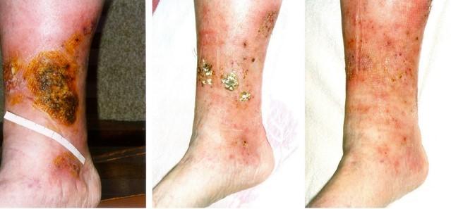
By Prof. Dr. med. Gerd Hoffmann, CC BY-SA 3.0 DE, via Wikimedia Commons
Note considerable eczema and early ulceration just above the medial malleolus. As well as the crusting of eczema, there are mottled pigmentary changes from haemosiderin.
During examination, look specifically for:
Skin changes - nature and severity. Look for:
The red, scaly or flaky skin of venous eczema. There may also be blisters and crusts on the surface.
Lipodermatosclerosis:
Hardened, tight, red or brown skin.
Usually affecting the inner aspect of the calf.
The subcutaneous tissues may become hard and depressed ('inverted champagne bottle' leg if the damage is circumferential).
It can present acutely and be wrongly diagnosed as cellulitis (or phlebitis).
Atrophie blanche:
Star-shaped, white (ivory), depressed, and atrophic scars surrounded by pigmentation.
Frequently found where an ulcer has healed.
Venous ulceration.
Dependent oedema.
Presence or absence of foot pulses.
Varicose veins. Note the location and severity of any varicose veins. They may not be apparent until the patient stands.
The classic tourniquet tests are no longer used by vascular specialists, as they are inaccurate and ultrasonography is now available.4 5
Continue reading below
Investigation
The history and clinical examination will not always indicate the nature and extent of the underlying abnormality.
Consider measuring the ankle brachial pressure index (ABPI) using a Doppler machine if use of compression stockings is being considered. Some consider this unnecessary if the foot pulses are easily palpable, and the person has no symptoms of arterial disease.1
Duplex ultrasound may be used where relevant to confirm varicose veins and assess extent.4
Primary care treatment and management of varicose eczema1
General advice
Avoid injury to the skin (eg, against furniture). This may very easily lead to ulceration.
Elevate the legs when sitting.
Keep physically active. Encourage regular walks.
Basic skin care
Advise regular use of emollient.
Treat symptom flares with a topical steroid (usually of moderate strength). The skin is usually dry and may be ulcerated and so an ointment may be preferable to a cream.
Try to avoid potential skin sensitisers during management.6
Below-knee compression hosiery
Provided that there is no arterial insufficiency, below-knee compression stockings should be worn. As above, Doppler testing may be required to ascertain arterial competence first. People are often reluctant to use them for a number of reasons which include discomfort, difficulty putting them on and cosmetic appearance.7 Stockings come in three grades of pressure:
Class 2 (medium) stockings, which are suitable for most people.
Class 1 (light) stocking if the person cannot tolerate a class 2 stocking.
Class 3 (strong) stockings, which may be necessary if the response to a class 2 stocking is inadequate (however, many people find these difficult to tolerate).
If ABPI is less than 0.3 or greater than 1.3, support stockings should not be worn. If ABPI is between 0.5 and 0.8, only a class 1 stocking should be used.8
Poor response to the above
If there is poor response then consider:
Contact dermatitis (for example, to applied topical treatments or materials in compression stockings).
Flares of lipodermatosclerosis may require application of very potent topical steroids.
Secondary infection. This may need treatment with topical or usually oral antibiotics.
Herbal remedies
There is some evidence from Cochrane reviews that oral horse chestnut seed extract may be of benefit for symptoms of chronic venous insufficiency.9 Further trials are needed, however.
When to refer1
This condition is likely to require involvement of different disciplines. Do not be reluctant to use the expertise of other members of the primary healthcare team.
Refer according to any local policies.
When there are no local policies, consider referral when:
Varicose veins present with progressive skin changes or a history of ulceration. Referral is usually to a vascular surgeon.
There is significant peripheral arterial disease (Doppler-measured ABPI of less than 0.8). Again referral to a vascular surgeon is recommended.
There is inadequate control of skin disease with primary care management (above). Referral to a dermatologist is recommended.
There is suspected contact dermatitis. Contact allergic dermatitis to paste bandages and medicaments applied to leg ulcers is common. If suspected, either because the eczema does not heal up or because it has recently flared, refer the patient to a dermatologist for further management and patch testing.
When discussing referral, take into consideration factors such as general state of health and comorbidities.
Prognosis of varicose eczema
This is a chronic condition and takes a long time to heal.
Topical steroids should clear the eczema but the secondary pigmentary changes will persist.
Poor adherence to strategies such as support hosiery or bandages may make prognosis worse than it should be.
If ulceration occurs, it will be a slower resolution.
If there is arterial insufficiency, healing is poor.
Complications include cellulitis, ulceration and contact dermatitis.
Prevention
There may be scope for prevention of skin disease and other complications with:
Better management of varicose veins.
Better management of venous insufficiency.
Prevention of DVT - surgery, flights, etc.
Better DVT detection and management.
All of this might be achieved with good primary care and timely referral to the appropriate specialist.
Further reading and references
- Gloviczki P, Comerota AJ, Dalsing MC, et al; The care of patients with varicose veins and associated chronic venous diseases: clinical practice guidelines of the Society for Vascular Surgery and the American Venous Forum. J Vasc Surg. 2011 May;53(5 Suppl):2S-48S. doi: 10.1016/j.jvs.2011.01.079.
- Barron GS, Jacob SE, Kirsner RS; Dermatologic complications of chronic venous disease: medical management and beyond. Ann Vasc Surg. 2007 Sep;21(5):652-62.
- Venous eczema and lipodermatosclerosis; NICE CKS, March 2022 (UK access only)
- Venous Eczema; DermNet
- Nazarko L; Diagnosis and treatment of venous eczema. Br J Community Nurs. 2009 May;14(5):188-94.
- Marsden G, Perry M, Kelley K, et al; Diagnosis and management of varicose veins in the legs: summary of NICE guidance. BMJ. 2013 Jul 24;347:f4279. doi: 10.1136/bmj.f4279.
- Campbell B; Varicose veins and their management. BMJ. 2006 Aug 5;333(7562):287-92.
- Beldon P; Avoiding allergic contact dermatitis in patients with venous leg ulcers. Br J Community Nurs. 2006 Mar;11(3):S6, S8, S10-2.
- Raju S, Hollis K, Neglen P; Use of compression stockings in chronic venous disease: patient compliance and efficacy. Ann Vasc Surg. 2007 Nov;21(6):790-5.
- Compression stockings; NICE CKS, May 2022 (UK access only)
- Pittler MH, Ernst E; Horse chestnut seed extract for chronic venous insufficiency. Cochrane Database Syst Rev. 2012 Nov 14;11:CD003230. doi: 10.1002/14651858.CD003230.pub4.
Continue reading below
Article history
The information on this page is written and peer reviewed by qualified clinicians.
Next review due: 17 Nov 2027
18 Nov 2022 | Latest version

Ask, share, connect.
Browse discussions, ask questions, and share experiences across hundreds of health topics.

Feeling unwell?
Assess your symptoms online for free