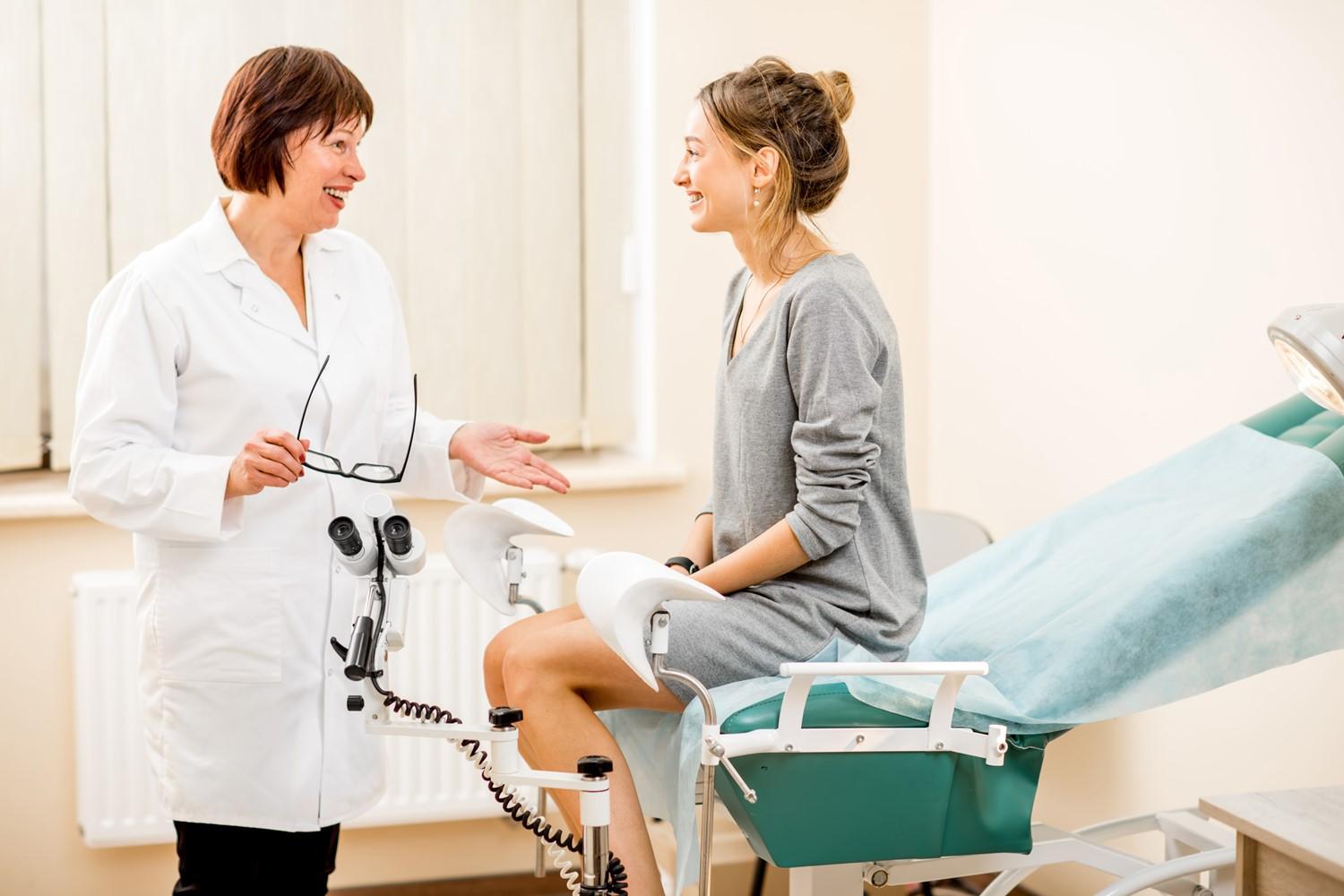
The female reproductive system
Peer reviewed by Dr Helen Huins, MRCGPLast updated by Dr Mary Harding, MRCGPLast updated 31 May 2018
Meets Patient’s editorial guidelines
- DownloadDownload
- Share
- Language
- Discussion
The organs, structures, and functions of the female reproductive system give women the ability to produce a baby. They do this by producing eggs, in monthly cycles known as the menstrual cycle, to be fertilised by sperm from a man. Other structures such as the breasts give the mother the ability to feed and nourish a baby after birth.
In this article:
Continue reading below
What are the main parts of the female reproductive system?
Uterus and ovaries
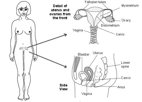
The organs of the female reproductive system are found both inside and outside of the female body. The organs inside the body are in the pelvis, which is the lowest part of the body cavity above the legs.
The internal female reproductive organs include:
Vagina - the area between the lower part of the womb (the cervix) and the outside of the body. The vagina receives the penis during sexual intercourse and is a passageway for childbirth.
Womb (uterus) - a hollow, pear-shaped organ that is the home to a developing fetus. The uterus is divided into two parts: the cervix, which is the lower part that opens into the vagina, and the main body of the uterus, called the corpus. The corpus can easily expand to hold a developing baby. A channel through the cervix allows sperm to enter and menstrual blood to exit.
Ovaries - small, oval-shaped glands that are located on either side of the uterus. The ovaries produce eggs (ova - an ovum is one egg, ova means multiple eggs.) The ovaries also produce the main female sex hormones which are released into the bloodstream.
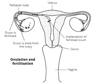
Uterine (Fallopian) tubes - narrow tubes that are attached to the upper part of the uterus. They serve as tunnels for the ova to travel from the ovaries to the uterus. The fertilisation of an egg by a sperm (conception) normally occurs in the uterine tubes. The fertilised egg then moves to the uterus, where it implants into the lining of the uterine wall.
Ovulation and fertilisation
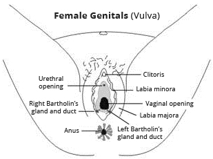
The outside (external) structures of the female reproductive system are grouped together in an area called the vulva. They are located just outside the opening of the vagina. This includes structures such as the labia, the clitoris and a number of glands. The breasts can also be considered part of the female reproductive system and are located on the chest.
The vulva consists of:
The labia - skin flaps or folds on either side of the opening of the vagina. There are two layers of these skin folds. The outer ones are called the labia majora and are covered with pubic hair after puberty. The inner folds do not have hair and are called the labia minora.
The mons pubis - the fatty bulge above the labia which is covered with hair after puberty.
The vaginal opening (meatus) - the entrance to the vagina.
The urethral opening (meatus) - the end of the tube which carries urine from the bladder to the outside (urethra).
The clitoris - a lump of tissue at the top of the labia. This becomes full of blood during sexual excitement (like the penis in the man but much much smaller). The clitoris is very sensitive and is the main source of female sexual pleasure.
The Bartholin's glands (or vestibular glands) - glands on either side of the opening of the vagina. These produce a sticky substance to moisten (lubricate) the vagina for sexual intercourse.
Female Genitals
What is the purpose of the female reproductive system do?
Back to contentsThe main function of the female reproductive system is to produce eggs (ova) to be fertilised, and to provide the space and conditions to allow a baby to develop. In order for this to happen, the female reproductive system also has the structures necessary to allow sperm from a man to meet the ova of a woman.
The female reproductive system makes its own hormones that help to control a woman's monthly cycle. These hormones cause ova to develop and be released in a monthly cycle. This process is called ovulation. If one of these ova is fertilised by a male sperm, this leads to pregnancy. The hormones also create the right conditions in the womb (uterus) for the fetus to develop, and block ovulation during pregnancy.
Continue reading below
How does the female reproductive system work?
Back to contentsThe activity of the female reproductive system is controlled by hormones released both by the brain and by the ovaries. The combination of all these hormones gives women their menstrual cycle.
The length of the menstrual (or reproductive) cycle is usually between 24-35 days. During this time an egg (ovum) is developed and matured. At the same time the lining of the womb (uterus) is prepared to receive a fertilised egg. If a fertilised egg is not implanted into the uterus, the lining of the uterus is shed and is expelled from the body. This is the bleeding known as having a period, or menstruation. Traditionally, the first day of bleeding is known as day one of the menstrual cycle. The key event in the cycle is ovulation. This is the release of a mature egg from one of the ovaries. This usually takes place around the 14th day of a 28-day cycle.
What hormones are released during the menstrual cycle?
Back to contentsThere are five main hormones that control the menstrual cycle. Three are produced in the brain, while the other two are made in the ovaries.
Gonadotrophin-releasing hormone (GnRH) is made by a part of the brain called the hypothalamus. GnRH travels to another part of the brain where it controls the release of follicle-stimulating hormone (FSH) and luteinising hormone (LH).
FSH is released by a part of the brain called the anterior pituitary. FSH is carried by the bloodstream to the ovaries. Here it stimulates the immature eggs (ova) to start growing.
LH is also released by the anterior pituitary and travels to the ovaries. LH triggers ovulation and encourages the formation of a special group of cells called the corpus luteum.
Oestrogen is produced by the growing ova and by the corpus luteum. In moderate amounts oestrogen helps to control the levels of GnRH, FSH and LH. This helps to prevent the development of too many ova. Oestrogen also helps to develop and maintain many of the female reproductive structures.
Progesterone is mainly released by the corpus luteum. It works with oestrogen to thicken the lining of the uterus ready for the implantation of a fertilised ovum. It also helps to prepare the breasts for releasing milk. High levels of progesterone control the levels of GnRH, FSH and LH.
During the last few days of the menstrual cycle, around 20 small immature ova begin to develop in the ovaries. This continues throughout the menstrual cycle. FSH and LH encourage the growth of these ova. As they grow, the ova also start to release increasing amounts of oestrogen. The amount of oestrogen produced reduces the amount of FSH released. This helps to prevent too many ova growing at the same time. Eventually one ovum outgrows the rest.
While this is happening in the ovaries, the oestrogen produced also causes a thickening of the lining of the uterus.
Continue reading below
What is ovulation?
Back to contentsOvulation is the stage in the menstrual cycle when the mature ovum is released from the ovaries into the Fallopian tubes. By this point in the cycle, levels of oestrogen are high. Previously, medium levels of oestrogen reduced the amount of FSH and LH released. Now this high level of oestrogen is the signal for more FSH and LH to be released. LH causes the ovum to burst through the outer layer of the ovary. Usually the ovum is then swept into the uterine tubes.
When the ovum leaves the ovary, the cells left behind become the corpus luteum. This special group of cells is capable of producing several different hormones, including progesterone and oestrogen. These hormones encourage the growth and maturation of the lining of the uterus.
What happens next depends on whether the ovum is fertilised by sperm. If the ovum is fertilised, the corpus luteum continues to produce hormones. Another hormone called human chorionic gonadotrophin (hCG) stops the corpus luteum from breaking down. The cells covering the embryo produce hCG. It is the hormone detected in pregnancy tests.
If the ovum is not fertilised, the corpus luteum can only live for a further two weeks. As it begins to break down, it releases fewer of its hormones. As the levels of progesterone and oestrogen go down, they no longer control the levels of GnRH, FSH and LH. So, these hormones increase and new ova begin to develop - the start of a new menstrual cycle. In the uterus the decrease in progesterone stimulates the release of chemicals that eventually cause the lining of the uterus to die off. This is the blood flow experienced during menstruation.
Some disorders of the female reproductive system
Back to contentsPatient picks for Women's sexual health

Sexual health
The most likely causes of pain during sex
Whilst it's not a topic many of us discuss, sex can sometimes prove a real pain for some women. In fact, around a third of younger and half of older women have experienced pain during sex, according to the Sexual Advice Association. As well as discomfort, painful sex can lead to problems in relationships, loss of intimacy and even depression. The good news is that many of these problems can be treated effectively by your GP or after a referral to a gynaecologist - so don't suffer in silence!
by Gillian Harvey

Sexual health
How to treat vaginismus
Vaginismus is when your vaginal muscles involuntarily tighten when you try to insert something into it. It can make having sex impossible or very painful. We find out what it’s like to live with the condition and how you can treat it to alleviate your symptoms.
by Sally Turner
Continue reading below
Article history
The information on this page is peer reviewed by qualified clinicians.
31 May 2018 | Latest version

Ask, share, connect.
Browse discussions, ask questions, and share experiences across hundreds of health topics.

Feeling unwell?
Assess your symptoms online for free
Sign up to the Patient newsletter
Your weekly dose of clear, trustworthy health advice - written to help you feel informed, confident and in control.
By subscribing you accept our Privacy Policy. You can unsubscribe at any time. We never sell your data.