Shoulder pain
Peer reviewed by Dr Toni Hazell, MRCGPLast updated by Dr Doug McKechnie, MRCGPLast updated 15 Oct 2024
Meets Patient’s editorial guidelines
- DownloadDownload
- Share
- Language
- Discussion
- Audio Version
Medical Professionals
Professional Reference articles are designed for health professionals to use. They are written by UK doctors and based on research evidence, UK and European Guidelines. You may find the Shoulder pain article more useful, or one of our other health articles.
In this article:
Continue reading below
What is shoulder pain?
Shoulder pain is a common symptom in primary care. It can be due to an intrinsic shoulder problem like calcific tendonitis, but pain can also be referred from other structures such as the neck, diaphragm or the heart.
Common shoulder problems share overlapping clinical features. When assessing shoulder pain, it is important to look for any 'red flags' that mean investigation and diagnosis need a more focused or urgent approach.
Anatomy of the shoulder joint
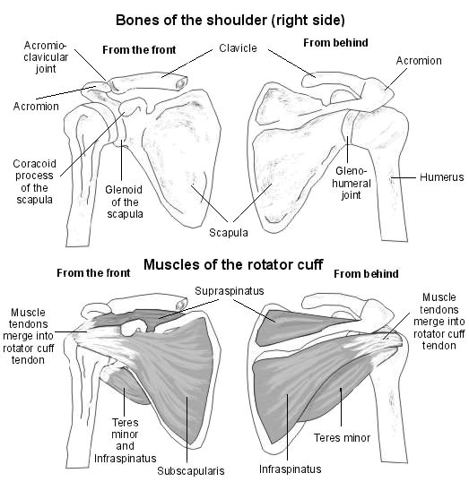
Back to contentsThe humerus, glenoid, scapula, acromion, clavicle and surrounding soft tissues make up the shoulder. There are three significant articulations: the sternoclavicular joint, the acromioclavicular joint and the glenohumeral joint. The glenohumeral joint is the most commonly dislocated major joint in the body.
Ligaments and surrounding musculature, including the rotator cuff muscles, contribute to shoulder joint stability. The rotator cuff is composed of the four muscles: supraspinatus, infraspinatus, teres minor and subscapularis that interlock to function as one unit. These muscles help with internal and external rotation of the shoulder and importantly depress the humeral head against the glenoid as the arm is elevated.
The tendons join together to form one tendon, the rotator cuff tendon. This passes through the subacromial space. The subacromial bursa, which has a large number of pain sensors, fills the space between the acromion and the rotator cuff tendon.1
Rotator cuff
Continue reading below
How common is shoulder pain? (Epidemiology)2
Back to contentsShoulder pain is the third most common cause of musculoskeletal consultation in primary care.
1% of adults with new shoulder pain consult their GP each year.
Self-reported prevalence of shoulder pain is between 16% and 26%.
Risk factors for shoulder pain
Back to contentsPhysical factors related to occupation including repetitive movements and exposure to vibration from machine tools.
Psychosocial factors related to work may also be risk factors for shoulder pain, including stress, job pressure, social support and job satisfaction.
Athletes whose sports involve overhead activities, or high-impact contact sports, are prone to shoulder pain.
Occupations particularly prone to shoulder pain syndromes include: cashiers, garment makers, bricklayers/construction workers, pneumatic tool operators, welders, meat/food-processing workers, hairdressers, plasterers, painters and decorators, assembly/production line workers, and workers using keyboards for long periods - eg, IT, secretarial.
Continue reading below
Shoulder pain causes23
Back to contentsPatients presenting in primary care often have a combination of different shoulder problems.
Intrinsic shoulder pain:
Subacromial pain syndrome (SAPS) - the most common cause of shoulder pain in primary care. This is a general term which includes:
Shoulder impingement.
Subacromial bursitis.
Rotator cuff tears and tendinopathy.
Calcific tendinitis.
Biceps tendinitis.
Glenohumeral disorders: adhesive capsulitis ('frozen shoulder'), arthritis.
Infection (rare).
Traumatic dislocation and fractures.
Shoulder instability - associated with hypermobility, including subluxation or dislocation (see also the separate article Shoulder dislocation).
Extrinsic shoulder pain:
Referred pain: neck pain, myocardial ischaemia, referred diaphragmatic pain (eg, gallbladder disease, subphrenic abscess).
Malignancy: apical lung cancers, metastases.
Rotator cuff disorders
The term subacromial pain (synonyms: subacromial impingement; impingement syndrome; rotator cuff syndrome; supraspinatus tendonitis; rotator cuff tendinopathy; painful arc syndrome) refers to all rotator cuff lesions, including all stages of tendon disease from early degeneration through to complete tears.
Most often present in patients aged 35-75 years.
Subacromial impingement is the most common source of shoulder pain:
There may be a history of heavy lifting or repetitive movements, especially above shoulder level. However, it often occurs in the non-dominant arm and in non-manual workers.
On examination there may be muscle wasting with pain on movements and a partial restriction of active movements (passive movements are full but painful).
A painful arc (between 70-120° of active abduction) is not specific or sensitive but increases the likelihood of a rotator cuff disorder.
A rotator cuff tear:
Usually follows trauma in young people. It is usually atraumatic in elderly people and caused by attrition from bony spurs on the undersurface of the acromion or intrinsic degeneration of the cuff, possibly.
Partial tears may be difficult to differentiate from rotator cuff tendinopathy on examination.
The drop arm test (see 'Examination', below) may be used to detect a massive tear.
Calcific tendonitis:4
Crystalline calcium phosphate is deposited in the rotator cuff tendon.
The cause is not known. It is more common in women (70% of cases) and affects people aged 30-60.
It is a self-limiting condition as the calcium will eventually resorb but may take many years.
Glenohumeral disorders
Adhesive capsulitis most often presents between the ages of 40-65 years, whereas osteoarthritis is most common in those aged 60 years or older.
Adhesive capsulitis (frozen shoulder) and arthritis often present with a history of non-adhesive capsulitis symptoms, cause deep joint pain and restrict activities such as putting on a jacket - because of impaired external rotation.
Adhesive capsulitis is more common in people with diabetes and may also occur after prolonged immobilisation.
There is usually generalised shoulder pain and a restriction of passive and active movements.
Acromioclavicular disorders
See also the separate article Acromioclavicular joint problems.
They are usually caused by trauma or osteoarthritis.
Pain and tenderness are localised to the acromioclavicular joint and there is a restriction of passive, horizontal movement of the arm across the body when the elbow is extended.
Obvious deformity after injury suggests a significant tear of the acromioclavicular ligament .
Acromioclavicular osteoarthritis may cause subacromial impingement.
Referred neck pain
See also the separate article Neck pain (cervicalgia) and torticollis.
Typically, this presents with pain and tenderness of the lower neck and suprascapular area, with pain referred to the shoulder and upper arm.
There may be a restriction of shoulder movement and movement of the neck and shoulder may reproduce more generalised upper back, neck and shoulder pain.
There may also be upper limb paraesthesia.
Shoulder pain assessment2
Back to contentsWhen assessing shoulder pain, take a history and perform an examination with these questions in mind:
Is the pain arising from the shoulder, neck or elsewhere?
Are there any 'red flag' symptoms/signs?
Is the pain localised to the acromioclavicular joint: the 'pointing sign'? If yes, there is acromioclavicular joint disease.
Is there global pain and restriction of all active and passive movements? If yes, this suggests glenohumeral joint disorder (either 'frozen shoulder' or arthritis).
Does the patient show a broad area of pain: the 'grasping sign' suggestive of subacromial pain?
History
Points to cover in the history include:2
The nature of the pain including:
How the pain started.
Any specific injury.
Whether it is acute or chronic.
Any impact on function/activities of daily living.
Whether the pain is on the side of the dominant hand.
Whether there is pain at rest or on movement.
Whether there is night pain that affects sleep.
Any associated pain - for example, neck, chest or other upper limb or joint pain.
Any history of shoulder pain/instability/dislocation.
The patient's occupation.
The patient's sporting activities.
Any signs or symptoms of systemic illness.
Past medical history (particularly any history of diabetes, coronary heart disease, cancer).
Drug history and adverse drug reactions.
Examination2 5
See the separate article Shoulder examination for further information.
There are over 100 specific 'Orthopaedic Special Tests' to detect shoulder pathology but few are sensitive or specific enough to be diagnostically discriminatory.6
Examine the neck, axilla and chest wall.
Examine the cervical spine and assess range of motion.
Inspect from the front, side, and behind for muscle wasting, swelling and deformity, or for bruising.
Palpate the sternoclavicular, acromioclavicular and glenohumeral joints. Look for tenderness, swelling, warmth and crepitus.
As an initial screening test, ask the person to place the palms of their hands at the base of the neck with elbows pointing laterally and then to put their arms down and try to put the back of the hands between the shoulder blades. However, be aware that this also involves joints other than the shoulder (ie elbow, wrist).
Assess the power, stability and range of motion (active, passive and resisted) in both shoulders.
Look for a painful arc (pain between 70-120° of abduction).
Test passive external rotation (reduced in 'frozen shoulder'). With the elbow held into the side, turn the arm outwards as far as possible.
Perform the 'drop arm test': passively abduct the patient's shoulder. Then ask the patient to lower the abducted arm slowly to the waist. This can identify a massive rotator cuff tear. They may be able to lower the arm slowly to 90° because this uses mostly the deltoid muscle but, below 90°, the arm will drop to the side.
Perform the 'cross-arm test': this isolates the acromioclavicular joint. Ask the patient to raise the arm to 90° straight in front of them. Then ask the patient to adduct the arm across the chest. If there is an acromioclavicular joint problem, there will be pain in the area of the joint.
Diagnosing shoulder pain (investigations)
Back to contentsBlood tests including FBC, ESR/CRP and radiology such as CXR are generally only necessary if there are 'red flag' symptoms/signs.2
Ultrasonography is useful to evaluate the integrity of the rotator cuff, if rotator cuff tears are suspected. Note however that asymptomatic full-thickness degenerative tears of the rotator cuff are common in older people.3
Plain X-rays are of relatively limited use in primary care. They are useful if a fracture or dislocation is suspected, and can identify osteoarthritis in the acromioclavicular and glenohumeral joints. However, asymptomatic radiological osteoarthritis of these joints is common; X-ray imaging is only indicated if there is a clinical suspicion of one of these diagnoses.3
Magnetic resonance arthrogram is useful in shoulder instability.7
If referred neck pain is suspected then cervical spine imaging, such as MRI, may be helpful but the diagnosis is usually clinical.
Shoulder pain treatment and management
Back to contentsSee also the separate article Shoulder joint replacements.
There is a lack of well-designed clinical trials in the management of shoulder disorders. Management in primary care is usually conservative: reduce or avoid overhead activities; attention to any contributing factors; medication for pain relief, including corticosteroid injection. If symptoms don't settle quickly or are severe initially, physiotherapy focused on the specific cause is indicated.8
Subacromial pain syndrome:3
Offer analgesia; ideally an oral non-steroidal anti-inflammatory drug (NSAID), or paracetamol and/or if unsuitable.
Refer to physiotherapy with the goal of optimising shoulder function, using an evidence-based rehabilitation protocol.9
Consider a subacromial corticosteroid injection if the person has limited function because of pain and is therefore unable to perform strengthening and stabilising exercises. Subacromial corticosteroid injections may provide short term benefit (at eight weeks), but no long-term benefit (at 12 months).10 See the separate Joint injection and aspiration article.
Do not give a corticosteroid injection if:
The person has previously received a corticosteroid injection from an experienced practitioner with minimal or no benefit.
The person has already had three or more injections in the same shoulder in the previous year.
There is a suspected significant rotator cuff tear.
There is any contra-indication to corticosteroid injection (eg, infection, osteomyelitis).
Rotator cuff tears:
Acute traumatic rotator cuff tears should be referred urgently to orthopaedics;11these patients may be a candidate for urgent operative repair.3
Degenerative or chronic rotator cuff tears may be managed initially with analgesia, physiotherapy, and/or steroid injections, with potential onward referral if these fail to control symptoms.12
Surgical treatment usually involves arthroscopic rotator cuff tendon repair.
Calcific tendonitis:4
When calcific tendonitis is symptomatic, it may present as chronic, relatively mild pain in the shoulder, with sporadic episodes of severe, acute pain radiating down the arm or to the neck.
The calcium deposits cause a chemical irritant inflammatory reaction. There is also an increase in pressure in the tendon, which is turn leads to malfunction of the rotator cuff and subacromial pain.
Treatment for calcific tendonitis includes NSAIDs, corticosteroids, physiotherapy, aspiration or lavage. For patients refractory to these treatments, open or arthroscopic shoulder surgery may be offered to excise the deposit.
Extracorporeal shock wave lithotripsy is no longer recommended by the National Institute for Health and Care Excellence (NICE).13
Glenohumeral disorders: see the separate article Frozen shoulder.
Glucocorticoid injection appears to be more effective in the short term than physiotherapy and exercises.14
Acromioclavicular disease (see the separate Acromioclavicular joint problems article):
Acromioclavicular injury usually responds to rest and simple analgesia, unless there is significant disruption of the joint, in which case orthopaedic referral is necessary.15
Consider providing a sling for 5-7 days if an acromioclavicular joint injury is suspected.
Consider referring to physiotherapy after 4-6 weeks if the person responds poorly to rest and analgesia.
Degeneration of the humeral head:16
The humeral head may degenerate as a result of a range of conditions - eg, osteoarthritis, rheumatoid arthritis or avascular necrosis. The whole or only part of the articular surface of the humeral head may be affected.
Conservative treatment includes physiotherapy, pain relief, topical or oral NSAIDs and corticosteroid injections.
Patients who do not respond to conservative treatments may need surgery, which involves either shoulder arthroplasty using a stemmed humeral head prosthesis, or fusion of the joint.
Shoulder resurfacing arthroplasty replaces only the damaged joint surfaces, with minimal bone resection and is recommended by NICE as a surgical option.
Muscle strains:
Muscle strains of the shoulder are very common, including trapezius and rhomboid strains.
Treatment of muscle strains includes rest, ice/heat, compression and elevation (sitting up) as well as massage, non-steroidal anti-inflammatory drugs (topical or oral) and further analgesia as required.
Severe or persistent strains may require physiotherapy.
There is no good-quality evidence to say whether acupuncture works to treat shoulder pain of any cause or if it is harmful.17
Criteria for referral to secondary care 53
Back to contentsEmergency same day assessment if:
Suspected joint infection (red skin, fever, swollen joint or systemically unwell).
Unreduced dislocation (trauma, epileptic fit or electric shock leading to abnormal shoulder shape and loss of rotation).
Acute trauma, depending on clinical judgement.
Acute neurological injury or pathology - a sudden serious motor or sensory deficit of the arm.
Urgent if any red flags are identified:
Trauma, pain and weakness, or sudden loss of ability to actively raise the arm (with or without trauma): suspect acute rotator cuff tear.
A new, unexplained shoulder mass or swelling: suspect malignancy.
New symptoms of inflammation in several joints: suspect inflammatory arthritis.
Consider urgent investigations and/or referral to secondary care if:
Systemic symptoms - eg, fever, night sweats, weight loss or new respiratory symptoms.
Undiagnosed severe shoulder pain or severe restriction of movement.
Refer early to secondary care if:
Recurrent shoulder instability.
Severe post-traumatic pain.
Pain is having a significant impact - eg, work or sport activities.
Refer if pain and function are not improving following conservative treatment for three months.
Consider referral to a specialised musculoskeletal clinic - for example, for provision of physiotherapy or corticosteroid injection.
Shoulder pain prognosis8
Back to contentsThe prognosis of chronic shoulder pain depends on the underlying cause.
Increasing age, female sex, symptoms of gradual onset, prolonged symptoms, severe or recurrent symptoms and associated neck pain are associated with a worse outcome.2
Recovery in shoulder pain is generally slow. Studies have shown complete recovery at one month in 23% of patients and at 18 months in 59% of patients.
Further reading and references
- Shoulder Disorders; Wheeless' Textbook of Orthopaedics
- Prof L Funk; Shoulder and Elbow Information, ShoulderDoc
- Mitchell C, Adebajo A, Hay E, et al; Shoulder pain: diagnosis and management in primary care. BMJ. 2005 Nov 12;331(7525):1124-8.
- Murphy RJ, Bintcliffe F; Ask the expert: assessment of shoulder pain in primary care. BMJ. 2023 Jul 7;382:1255. doi: 10.1136/bmj.p1255.
- Serafini G, Sconfienza LM, Lacelli F, et al; Rotator cuff calcific tendonitis: short-term and 10-year outcomes after two-needle us-guided percutaneous treatment--nonrandomized controlled trial. Radiology. 2009 Jul;252(1):157-64. doi: 10.1148/radiol.2521081816.
- Shoulder pain; NICE CKS, November 2022 (UK access only)
- Hegedus EJ, Goode A, Campbell S, et al; Physical examination tests of the shoulder: a systematic review with meta-analysis of individual tests. Br J Sports Med. 2008 Feb;42(2):80-92; discussion 92. Epub 2007 Aug 24.
- Burbank KM, Stevenson JH, Czarnecki GR, et al; Chronic shoulder pain: part I. Evaluation and diagnosis. Am Fam Physician. 2008 Feb 15;77(4):453-60.
- Burbank KM, Stevenson JH, Czarnecki GR, et al; Chronic shoulder pain: part II. Treatment. Am Fam Physician. 2008 Feb 15;77(4):493-7.
- Kuhn JE; Exercise in the treatment of rotator cuff impingement: a systematic review and a synthesized evidence-based rehabilitation protocol. J Shoulder Elbow Surg. 2009 Jan-Feb;18(1):138-60. doi: 10.1016/j.jse.2008.06.004. Epub 2008 Oct 2.
- Hopewell S, Keene DJ, Marian IR, et al; Progressive exercise compared with best practice advice, with or without corticosteroid injection, for the treatment of patients with rotator cuff disorders (GRASP): a multicentre, pragmatic, 2 x 2 factorial, randomised controlled trial. Lancet. 2021 Jul 31;398(10298):416-428. doi: 10.1016/S0140-6736(21)00846-1. Epub 2021 Jul 12.
- Craig R, Holt T, Rees JL; Acute rotator cuff tears. BMJ. 2017 Dec 11;359:j5366. doi: 10.1136/bmj.j5366.
- Mathiasen R, Hogrefe C; Evaluation and Management of Rotator Cuff Tears: a Primary Care Perspective. Curr Rev Musculoskelet Med. 2018 Mar;11(1):72-76. doi: 10.1007/s12178-018-9471-6.
- Extracorporeal shockwave therapy for calcific tendinopathy in the shoulder. Interventional procedures guidance [IPG742]. National Institute for Health and Care Excellence, 9 November 2022.
- Page MJ, Green S, Kramer S, et al; Manual therapy and exercise for adhesive capsulitis (frozen shoulder). Cochrane Database Syst Rev. 2014 Aug 26;8:CD011275. doi: 10.1002/14651858.CD011275.
- Simovitch R, Sanders B, Ozbaydar M, et al; Acromioclavicular joint injuries: diagnosis and management. J Am Acad Orthop Surg. 2009 Apr;17(4):207-19.
- Shoulder resurfacing arthroplasty; NICE Interventional Procedure Guidance, July 2010
- Green S, Buchbinder R, Hetrick S; Acupuncture for shoulder pain. Cochrane Database Syst Rev. 2005 Apr 18;(2):CD005319.
Continue reading below
Article history
The information on this page is written and peer reviewed by qualified clinicians.
Next review due: 14 Oct 2027
15 Oct 2024 | Latest version

Ask, share, connect.
Browse discussions, ask questions, and share experiences across hundreds of health topics.

Feeling unwell?
Assess your symptoms online for free