Astigmatism
Peer reviewed by Dr Toni Hazell, MRCGPLast updated by Dr Doug McKechnie, MRCGPLast updated 15 Nov 2023
Meets Patient’s editorial guidelines
- DownloadDownload
- Share
- Language
- Discussion
Astigmatism is a common eye condition that affects the shape of the cornea or the lens of the eye. The cornea and lens are typically smooth and round in shape, but in people with astigmatism, they are irregularly shaped.
The main symptom of astigmatism is blurred vision. It occurs because the cornea at the front of the eye is unevenly curved. Vision problems, such as astigmatism, are also known as refractive errors. Astigmatism is a common condition that can be corrected by glasses or contact lenses, or cured with laser eye surgery.
In this article:
Video picks for Eye conditions
Continue reading below
What is astigmatism?
Astigmatism

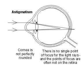
Astigmatism is a type of visual problem called a 'refractive error', meaning there is a problem in the eye that prevents the incoming light rays from being focused correctly. The shape of the cornea at the front of the eye is not perfectly rounded but is curved, a little like a rugby ball. When this curve is too great, or curves in the wrong direction, astigmatism occurs.
Light rays coming through the cornea and lens are not focused on to one sharp spot on the retina but are spread. This lack of 'point focus' means that images received by the brain are blurred.
Astigmatism is a bit more complicated than either short sight (myopia) or long sight (hypermetropia), as there is a problem with the focus of light in two different directions (both depth-ways and sideways).
However, like the other refractive errors, the end result is the same: there is blurring of vision which can impair eyesight. The two eyes are usually not the same (ie if both eyes have astigmatism they may not be affected in the same line of vision or to the same degree).
If the condition is mild, the brain will compensate for the difference between the two eyes, although this can cause eye strain and headaches. Although astigmatism is very common , it does not always cause a problem.
Astigmatism symptoms
Back to contentsFor most people with astigmatism, it is a very mild, minor problem which may not even be noticed. However, with more advanced astigmatism, there can be a variety of symptoms including:
Blurred vision.
Light sensitivity (photophobia).
Eye strain and fatigue (especially after long periods of concentration, such as when using a computer).
Headaches.
Other symptoms can include:
Difficulty seeing one colour against another (contrast).
Distorted images, such as lines which lean to one side.
Double vision (with severe astigmatism).
Astigmatism often occurs alongside either short sight (myopia), long sight (hypermetropia) or age-related long sight (presbyopia). See the separate leaflets called Short-sightedness - Myopia, Long-sightedness - Hypermetropia and Age-related Long Sight (Presbyopia) for more details.
Continue reading below
What causes astigmatism?
Back to contentsThere are two main types of astigmatism:
Corneal astigmatism is when the cornea - the clear front part of the eye - is misshapen and not perfectly round.
Lenticular astigmatism is when the lens of the eye is misshapen.
Astigmatism is usually present at birth. However, it can result from:
An eye injury.
A scar.
An operation to the eye, particularly if the corneal surface is damaged.
Anything pressing persistently on the surface of the cornea (such as a large lump on the eyelid) which pushes it out of shape.
Preterm babies are at risk of refractive errors, including astigmatism. This may be because the cornea did not have enough time to develop properly in the womb.
Is astigmatism genetic?
Astigmatism can run in families.
Problems with the structure of the cornea can cause astigmatism. About 1 in 5 people with Down's syndrome have this problem and have significant astigmatism. Other corneal disorders develop throughout life. The most common of these is a condition called keratoconus. This can cause significant astigmatism, as well as short sight (myopia) and corneal scarring.
Complications with astigmatism
Back to contentsLazy eye
Astigmatism in only one eye may cause lazy eye (amblyopia) if present from birth. The affected eye does not 'learn' how to see because the brain ignores the signals it receives.
Amblyopia can be treated with eye patching if diagnosed early enough, before the vision pathways in the brain are fully developed. See the separate leaflet called Amblyopia (Lazy Eye) for more details.
Continue reading below
Astigmatism diagnosis
Back to contentsAstigmatism is diagnosed with an eye exam, usually by an optometrist. Tests done include:
An autorefractor test, which measures the shape and focus of the eyes.
Retinoscopy, which shows how a beam of light moves once it enters the eye and is reflected back.
The Jackson cross cylinder test, which involves looking through different lenses whilst the optometrist asks which one makes things look clearer. It can be used to fine-tune the correction needed for astigmatism.
Astigmatism treatment
Back to contentsIn many cases the symptoms of astigmatism are so mild that no treatment is needed. If vision is more significantly affected, glasses, contact lenses or surgery can correct the vision. Some treatment options include:
Glasses
The simplest, cheapest and safest way to correct a regular astigmatism is with glasses. The lenses of the glasses adjust the direction of the incoming light rays, correcting the uneven curve of the cornea.
There is an enormous choice of spectacle frames available, to suit all budgets and styles. However, an irregular astigmatism (where the surface of the eye is not only curved instead of perfectly rounded, but is also irregular or curved in multiple directions) cannot be corrected by a lens.
Contact lenses
These do the same job as glasses and are often the best option for astigmatism. Toric lenses are used. Contact lenses sit right on the surface of the eye. Many different types of contact lenses are available.
These may be soft lenses or rigid gas-permeable lenses. They can be daily disposable, extended wear, monthly disposable, or non-disposable. Your optician can advise which type is most suitable for your eyes and your prescription.
Contact lenses tend to be more expensive than glasses. They provide good all-round vision and do not mist over (for example, while doing sports or in hot environments).
They do, however, require more care and meticulous hygiene, and should not be worn during swimming, showering or sleeping. They are more suitable for older teenagers and adults, rather than very young children.
Laser eye surgery
Laser eye surgery is an option for some people to fully correct their astigmatism and any associated short or long sight. Generally, these operations are not available on the NHS.
Complete and permanent resolution of the astigmatism is possible in a number of people. Others have a significant improvement even though perfect vision is not achieved, and glasses or contact lenses may still be needed.
Many private companies advertise laser eye surgery. Before embarking upon this type of treatment you should do some research. You only have one pair of eyes and you need to find the best treatment for you. This may not be the cheapest.
Refractive surgery must be carried out in premises registered with the Care Quality Commission (CQC) in England, or the equivalent regulator in Scotland, Wales and Northern Ireland. It is important that you know your facts, including what the procedure involves, the failure rate, side-effects, the risk of complications, and the level of aftercare provided.
You should be given the opportunity to discuss these facts in advance with the surgeon who will be carrying out the procedure.
Types of laser eye surgery
Back to contentsSeveral types of laser eye surgery have been developed. These include LASIK® and surface laser treatments (PRK®, LASEK® and TransPRK®). They are all similar, in that they aim to reshape the cornea by using a laser to remove a very thin layer of corneal tissue.
The reshaping of the cornea allows the refraction error to be corrected. They also all have similar risks and benefits, and the main difference between them is the speed of recovery after surgery.
LASIK®
LASIK stands for Laser-Assisted In situ Keratomileusis. This is the most popular type of laser eye surgery.
The surgery is done with two lasers; the first laser creates a thin flap of cornea, which is moved aside to allow the second laser to reshape the cornea.
The flap is then replaced, and sticks by itself to the underlying cornea without the need for stitches. The flap serves as a natural bandage, keeping the eye comfortable as it heals and allowing healing to occur relatively quickly.
Vision recovery time is said to be around 24 hours. People may be able to return to work the day after LASIK surgery, but need to wait for at least one month before doing any contact sports.
Surface laser treatments (PRK®, LASEK® and TransPRK)
PRK stands for Photo-Refractive Keratectomy.
LASEK stands for LAser Sub-Epithelial Keratomileusis.
In these types of treatments, no flap is created but, instead, the very thin layer of cells at the surface of the eye (epithelium) is either removed or displaced, and a laser is applied directly to the cornea underneath to reshape it. The epithelium will grow back naturally but, while it is doing this, the eye will be very sore.
All surface layer treatments produce similar results, and the only difference between them is the way in which the thin surface layer (epithelium) is removed. In PRK and LASEK it is removed by the surgeon. In TransPRK it can be done by the laser as part of the reshaping treatment.
Visual recovery tends to be slower after surface laser treatments than after LASIK, and patients may take a week or longer to reach the driving standard. However, patients can return to contact sports sooner than after LASIK.
SMILE® (Small Incision Lenticule Extraction) is another type of surface laser treatment that was approved for use by the US FDA to treat people who have short-sightedness (myopia) plus a certain degree of astigmatism.
Side-effects of all laser surgery include:
Discomfort.
Blurry vision.
Glare and haloes around lights (particularly at night).
Red marks on the white of the eyes, caused by burst small blood vessels (subconjunctival haemorrhages).
These usually get better over time. Over-correction or under-correction of short-sightedness can also happen. Complications include eye infection and dry eyes. Permanent loss of vision is very rare; if this happens, around 1 in 5,000 people need a corneal transplant to restore their vision. Up to 1 in 10 patients may need additional surgery to get the best result.
Other techniques
Other treatments include replacement of the eye's natural lens with an artificial lens (intraocular lens) and corneal grafts (which are done in very severe or specialised cases of astigmatism).
How often do I need an eyesight test?
Back to contentsThe NHS recommends that most people should get their eyesight tested every two years. Children will routinely be offered eye checks at various stages from birth to school age.
People at higher risk of sight problems need more frequent eyesight checks. If you have diabetes, raised pressure in the eye (glaucoma), macular degeneration, or a family history of these conditions, you should check to see what your optician or doctor recommends about regular check-ups.
People over the age of 70 years and children who wear glasses may also need more frequent eye tests. You should get your eyes checked if you notice any changes in your vision.
Some opticians offer a home visiting service to carry out sight tests for people who are unable to get out and about.
What is a refractive error?
Back to contentsA refractive error is an eyesight problem. Refractive errors are a common reason for reduced level of eyesight (visual acuity).
Eye cross-section

Refraction refers to the bending of light, in this case by the eye, in order to focus it. A refractive error means that the eye cannot focus light on to the retina properly. There are four types of refractive error:
Short sight (myopia).
Long sight (hypermetropia).
Age-related long sight (presbyopia).
Astigmatism (a refractive error due to an unevenly curved cornea).
In order to understand refractive errors fully, it is useful to know how we see.
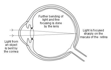
When we look at an object, light rays from the object pass through the eye to reach the retina. This causes nerve messages to be sent from the cells of the retina down the optic nerve to the vision centres in the brain. The brain processes the information it receives, resulting in an image that we can see.
Focusing the eye

Light rays come off an object in all directions, as they result from the light around us (for example, from the sun, moon and artificial light) bouncing back off the object. The part of this bounced light that comes into the eye from an object needs to be focused on a small area of the retina. If this doesn't happen, what we look at will be blurred.
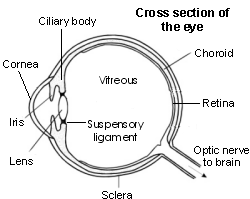
The cornea and lens have the job of focusing light. The cornea does most of the work, as it bends (refracts) the light rays which then go through the lens, which finely adjusts the focusing. The lens does this by changing its thickness. This is called accommodation. The lens is elastic and can become flatter or more rounded. The more rounded (convex) the lens, the more the light rays can be bent inwards.
The shape of the lens is varied by small muscles in the ciliary body. Tiny string-like structures called the suspensory ligaments are attached at one end to the lens and at the other to the ciliary body. This is a bit like a trampoline with the central bouncy bit being the lens, the suspensory ligaments being the springs and the ciliary muscles being the rim around the edge.
When the ciliary muscles in the ciliary body tighten, the suspensory ligaments slacken, causing the lens to become fatter. This happens for near objects. For looking at far objects, the ciliary muscle relaxes, making the suspensory ligaments tighten, and the lens thins out.
More bending (refraction) of the light rays is needed to focus on nearby objects, such as when reading. Less bending of light is needed to focus on objects far away.
Dr Mary Lowth is an author or the original author of this leaflet.
Patient picks for Eye conditions

Eye health
Myopia
The medical name for short-sightedness is myopia. Eyesight problems, such as myopia, are also known as refractive errors. Short-sightedness leads to blurred vision when looking at things at a distance, whilst close vision is usually normal. Short-sightedness is a very common problem that can be corrected by glasses, contact lenses, or laser eye surgery.
by Dr Philippa Vincent, MRCGP

Eye health
Contact lenses
Contact lenses are optical devices that sit over of the clear front surface of your eye (the cornea). They are most commonly worn as an alternative to spectacles to correct problems with your vision. They need to be looked after carefully.
by Dr Mary Elisabeth Lowth, FRCGP
Further reading and references
- Photorefractive (laser) surgery for the correction of refractive error; NICE Interventional procedures guidance, March 2006
- Professional Standards for Refractive Surgery; Royal College of Ophthalmologists (Dec 2021)
- Laser Vision Correction; Royal College of Ophthalmologists Patient Information Leaflet
- Chow SSW, Chow LLW, Lee CZ, et al; Astigmatism Correction Using SMILE. Asia Pac J Ophthalmol (Phila). 2019 Sep-Oct;8(5):391-396. doi: 10.1097/01.APO.0000580140.74826.f5.
- Keshav V, Henderson BA; Astigmatism Management with Intraocular Lens Surgery. Ophthalmology. 2021 Nov;128(11):e153-e163. doi: 10.1016/j.ophtha.2020.08.011. Epub 2020 Aug 13.
Continue reading below
Article history
The information on this page is written and peer reviewed by qualified clinicians.
Next review due: 13 Nov 2028
15 Nov 2023 | Latest version

Ask, share, connect.
Browse discussions, ask questions, and share experiences across hundreds of health topics.

Feeling unwell?
Assess your symptoms online for free
Sign up to the Patient newsletter
Your weekly dose of clear, trustworthy health advice - written to help you feel informed, confident and in control.
By subscribing you accept our Privacy Policy. You can unsubscribe at any time. We never sell your data.