PET scan
Peer reviewed by Dr Colin Tidy, MRCGPLast updated by Dr Rosalyn Adleman, MRCGPLast updated 26 Sept 2023
Meets Patient’s editorial guidelines
- DownloadDownload
- Share
- Language
- Discussion
- Audio Version
A PET scan create images which show where cells are particularly active in the body. It is most commonly used to diagnose and assess cancer.
Note: the information below is a general guide only. The arrangements, and the way tests are performed, may vary between different hospitals. Always follow the instructions given by your doctor or local hospital.
In this article:
Video picks for Imaging
Continue reading below
What is a PET scan?
PET stands for positron emission tomography. A PET scan produces three-dimensional, colour images of your body, using radionuclides. See the separate leaflet called Radionuclide Scan (Isotope Scan) for more information on radionuclides. PET scans show where cells are particularly active.
PET can be used to diagnose various medical conditions, or to find out more about how a condition is developing. It can also be used to measure how well treatment for a condition is working.
Increasingly, PET scans are being combined with another form of imaging such as magnetic resonance imaging (MRI) or computerised tomography (CT). By superimposing one scan on top of another, doctors can create very detailed pictures of the body.
What is a PET scan used for?
Back to contentsPET scanning is most commonly used in the diagnosis and assessment of cancer. However, it can be used to diagnose other conditions including Alzheimer's disease, epilepsy and heart disease. In cancer medicine, doctors may use the scan for the following reasons:
To detect a cancer. For example, to show whether a lump is cancer or not.
To see whether a cancer has spread to other parts of the body.
In decisions about the best treatment for a cancer.
To see how well treatment with cancer medication is working.
To show the difference between scar tissue and active cancerous cells.
If you have epilepsy, PET scanning may be used to assess which part of your brain is affected, and whether you are suitable for certain treatments.
In Alzheimer's disease, a PET scan can be used to provide a diagnosis of the condition.
PET scans of the heart can identify if parts of the heart have been scarred or damaged, and if it is working properly.
Continue reading below
What happens during a PET scan?
Back to contentsRadio-tracer injection
You will have a very small amount of a radio-tracer (nuclear medicine) injected into your arm, or breathe it in as a gas. It can take around 30-90 minutes to travel to the part of your body to be scanned. During this time, you will be asked to rest and to limit any movement or talking.
The scan
When you are ready for your scan, you will be taken to the scanning room. A PET scanner is a large machine with a round, doughnut-shaped hole in the middle, similar to a CT or MRI unit. Within this machine are multiple rings of detectors that record the emission of energy from the radio-tracer.
PET scanner
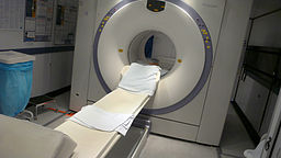
© By Liz West (Own work), via Wikimedia Commons
You will lie on a cushioned examination table, which is then moved into the large hole in the scanning machine so images of inside your body can be taken. During scanning you should stay as still as possible. It normally takes around 30-60 minutes to take a scan but it depends on which part of the body needs to be scanned.
Depending on which organ or tissue is being examined, additional tests involving other tracers or chemicals may be used, which could lengthen the procedure time. For example, if you are being examined for heart disease, you may undergo a PET scan both before and after exercising or before and after receiving medication that increases blood flow to the heart.
After the scan
When the scanning is completed, you may be asked to wait until the images are checked in case more images are needed. Occasionally, additional images are needed to clarify certain areas or structures. The need for additional images doesn't necessarily mean there was a problem or that something abnormal was found, and should not be a cause of concern for you. You will not be exposed to more radiation during this process.
What should I do to prepare for a PET scan?
Back to contentsYour healthcare professional will give you instructions on how to prepare for your scan that are specific to you. Normally you will be asked not to eat anything for 6 hours before the scan. This is because eating may change the distribution of the radio-tracer in your body and can lead to a poor-quality scan. If this happens, the scan may need to be repeated on another day.
So, following instructions regarding eating is very important. You may also be advised not to drink any caffeine in the 24 hours leading up to your scan. If you have diabetes you may receive special instructions about how to prepare for the scan.
You should advise your doctor if you are pregnant or think you may be pregnant. If you are breastfeeding you may be advised to express enough milk to feed your baby for the first six hours after the scan. This isn't because there will be radiation in the milk. It is because a mother shouldn't be holding the baby closely during the time the radiation is in her body. Your hospital should advise you on the precautions to take.
For other people, it is advisable that you do not have close contact with babies or young children until a few hours after your PET scan.
If you are claustrophobic or very nervous about the scan, you should discuss this with your doctor before the day of the scan. In some cases it is possible to have a mild sedative to prepare for your scan.
Continue reading below
What can I expect after a PET scan?
Back to contentsBecause the radiation exposure is low, you will not feel any effects and should be able to go home soon after your scan is completed. However, you should drink plenty of fluids afterwards to flush the radioactive drugs from your body. All traces of the radio-tracer should leave your body naturally around three hours after it has been given to you.
The scan will be reported by a consultant radiologist. The results will be sent to the doctor who ordered the scan. Usually results are available within a couple of weeks.
Are there any risks of the scan?
Back to contentsThe term 'radioactivity' may sound alarming. But, the radioactive chemicals used in PET scans are considered to be safe and they leave the body quickly in the urine. The dose of radiation that your body receives is very small.
If you are pregnant or breastfeeding, there may be a risk to your baby if you have a PET scan. This is because even small amounts of radiation can damage a baby. If you are pregnant or breastfeeding, it is important that you inform the staff at the hospital before the scan is carried out. They can advise you on the precautions to take.
Very rarely people have an allergic reaction to the radioactive-tracer used for the scan. This might cause sweating, itching or difficulty breathing. If you feel unwell during your scan, tell a member of staff immediately.
How does a PET scan work?
Back to contentsBefore a PET scan is carried out, a radioactive medicine is produced in a machine called a cyclotron. The medicine is then 'tagged' to a natural chemical such as glucose, water, or ammonia. This makes what is called a radio-tracer.
Once the radio-tracer is inside the body, it goes to parts of the body that use the chemical it has been tagged to. For example, a radioactive drug called fluorodeoxyglucose (FDG) is commonly used as a radio-tracer. It is a radioactive form of glucose. When inside the body it goes to the tissues that use glucose for energy.
A PET scan works by detecting the energy released by positrons. Positrons are tiny particles which are made as the radio-tracer is broken down inside your body. As positrons are broken down they create gamma rays. These gamma rays are detected by the scanner, which creates a three-dimensional image. The image can show how parts of your body work, by the way in which it breaks down the radio-tracer.
Different levels of positrons are shown as different colours and brightness on a PET image. Some parts of the body break down natural chemicals such as glucose quicker than others. A PET scan is particularly useful in detecting cancer because most cancers use more glucose than normal tissue uses. Areas of greater intensity, called 'hot spots', show where large amounts of the radio-tracer have built up. Less intense areas, or 'cold spots', indicate a smaller concentration of radio-tracer.
A radiologist is a specialist doctor who has training in interpreting images of the inside of the body. A radiologist will look at the images that a PET scan produces, and report the results to the doctor who is treating you.
Patient picks for Imaging

Tests and investigations
Cerebral angiography
Cerebral angiography is a test that uses X-rays and a special dye to create pictures of the blood vessels that supply the brain. Note: the information below is a general guide only. The arrangements, and the way tests are performed, may vary between different hospitals. Always follow the instructions given by your doctor or local hospital.
by Dr Mary Harding, MRCGP

Tests and investigations
X-ray test
X-ray tests show bones and certain other tissues.
by Dr Hayley Willacy, FRCGP
Further reading and references
- Positron Emission Tomography - Computed Tomography (PET/CT); RadiologyInfo.org
- Ehman EC, Johnson GB, Villanueva-Meyer JE, et al; PET/MRI: Where might it replace PET/CT? J Magn Reson Imaging. 2017 Nov;46(5):1247-1262. doi: 10.1002/jmri.25711. Epub 2017 Mar 30.
- Recommendations for cross-sectional imaging in cancer management; Royal College of Radiologists, 2022
- Masdeu JC; Neuroimaging of Diseases Causing Dementia. Neurol Clin. 2020 Feb;38(1):65-94. doi: 10.1016/j.ncl.2019.08.003. Epub 2019 Nov 7.
Continue reading below
Article history
The information on this page is written and peer reviewed by qualified clinicians.
Next review due: 24 Sept 2028
26 Sept 2023 | Latest version

Ask, share, connect.
Browse discussions, ask questions, and share experiences across hundreds of health topics.

Feeling unwell?
Assess your symptoms online for free
Sign up to the Patient newsletter
Your weekly dose of clear, trustworthy health advice - written to help you feel informed, confident and in control.
By subscribing you accept our Privacy Policy. You can unsubscribe at any time. We never sell your data.