Xanthelasma
Peer reviewed by Dr Toni Hazell, MRCGPLast updated by Dr Colin Tidy, MRCGPLast updated 24 May 2022
Meets Patient’s editorial guidelines
- DownloadDownload
- Share
- Language
- Discussion
- Audio Version
- Add to preferred sources on Google
Medical Professionals
Professional Reference articles are designed for health professionals to use. They are written by UK doctors and based on research evidence, UK and European Guidelines. You may find the Hyperlipidaemia article more useful, or one of our other health articles.
In this article:
Synonym: xanthelasma palpebrum
Continue reading below
What is xanthelasma?
The appearance of xanthelasma is of yellow flat plaques over the upper or lower eyelids, most often near the inner canthus. They represent areas of lipid-containing macrophages but the exact pathophysiology is not known. In other areas of the body the individual lesion would be called a xanthoma; xanthelasma is the most common xanthoma.1 2
Xanthelasma
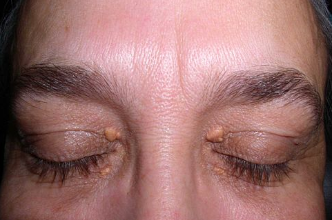
© By Klaus D. Peter, CC-BY-3.0, via Wikimedia Commons
Xanthelasma diagnosis
Back to contentsThis is usually not a problem, since colour and site are characteristic. They occur a little more often in women than in men and the peak incidence is in the fourth to fifth decades. Once the plaque is established, it tends to remain static in size or grow slowly; it does not regress. Generally, eyelid function remains unimpaired. Ptosis is rare.
Continue reading below
Differential diagnosis
Back to contentsSometimes syringomas and milia may be misdiagnosed as xanthelasma.
Syringomas are small papules on lower eyelids and are skin-coloured.
Large milial cysts are white and spherical.
Xanthomas in other areas may appear more orange-yellow.
The list of differentials for lipid disorders also needs to be considered.
Associated diseases
Back to contentsXanthelasma may represent a localised skin condition without any systemic abnormalities of lipoprotein metabolism or may be associated with an increase in the cholesterol-rich beta-lipoproteins (LDLs).3 See the separate Hyperlipidaemia article.
Some patients exhibiting xanthelasma have normal lipid levels but this is less common in younger patients. Although these patients are not at increased risk of carotid atherosclerosis, they are more commonly found to have other risk factors for cardiovascular disease - eg, a higher BMI, waist circumference and LDL-C levels.4
Continue reading below
Xanthelasma treatment and management
Back to contentsLesions are typically asymptomatic and treatment is sought for cosmetic purposes. Treatments include topical trichloroacetic acid, liquid nitrogen cryotherapy, and various lasers. Surgical excision has also been used.2
Patients should have their fasting lipid levels checked and those with hyperlipidaemia should have a formal cardiovascular risk assessment using appropriate charts, with measures for prevention of cardiovascular disease as indicated.
The lesions can be left alone unless the patient wishes them removed for cosmetic reasons (not usually available on the NHS).
Various options are available including surgical excision (with or without skin grafting for large lesions), chemical treatment, laser treatment and cryocautery.5 Full-thickness skin grafting obtained via blepharoplasty is available.6 Xanthelasmas may recur after any of these interventions.
Lipid-lowering medication and diet modification have a limited (if any) effect on these lesions.
Prognosis
Back to contentsThe condition itself is harmless. The prognosis depends on any association with underlying lipid abnormalities and cardiovascular risk.
When to refer
Back to contentsSurgical excision and cryocautery may be available in some primary care settings but it is likely that the other treatment options will require secondary care referral.
Exclusive updates for healthcare professionals
Stay informed with the latest clinical updates, professional insights, and evidence-based guidance. The Patient Pro newsletter curates essential content for healthcare professionals—delivered straight to your inbox.
By subscribing you accept our Privacy Policy. You can unsubscribe at any time. We never sell your data.
Further reading and references
- Xanthomata; Primary Care Dermatology Society.
- Xanthomas; DermNet NZ
- Laftah Z, Al-Niaimi F; Xanthelasma: An Update on Treatment Modalities. J Cutan Aesthet Surg. 2018 Jan-Mar;11(1):1-6. doi: 10.4103/JCAS.JCAS_56_17.
- Xanthelasma; DermIS (Dermatology Information System)
- Chan CC, Lin SJ, Hwang JJ, et al; Xanthelasma is not associated with increased risk of carotid atherosclerosis in normolipidaemia. Int J Clin Pract. 2008 Feb;62(2):221-7. Epub 2007 Nov 23.
- Elabjer BK, Busic M, Sekelj S, et al; Operative treatment of large periocular xanthelasma. Orbit. 2009;28(1):16-9.
- Kose R; Treatment of large xanthelasma palpebrarums with full-thickness skin grafts obtained by blepharoplasty. J Cutan Med Surg. 2013 May-Jun;17(3):197-200.
Continue reading below
About the authorView full bio

Dr Colin Tidy, MRCGP
General Practitioner, Medical Author
MBBS, MRCGP, MRCP (Paediatrics), DCH
Dr Colin Tidy is an NHS Doctor, based in Oxfordshire.
About the reviewerView full bio

Dr Toni Hazell, MRCGP
MBBS, BSc, MRCGP, DFSRH, Dip GU med, DRCOG, DCH (London, UK, 2000)
Dr. Toni Hazell qualified from St. Mary’s Hospital Medical School and did her VTS at Northwick Park Hospital.
Article history
The information on this page is written and peer reviewed by qualified clinicians.
Next review due: 8 Apr 2027
24 May 2022 | Latest version

Ask, share, connect.
Browse discussions, ask questions, and share experiences across hundreds of health topics.

Feeling unwell?
Assess your symptoms online for free