Vitiligo
Peer reviewed by Dr Doug McKechnie, MRCGPLast updated by Dr Colin Tidy, MRCGPLast updated 15 Dec 2022
Meets Patient’s editorial guidelines
- DownloadDownload
- Share
- Language
- Discussion
- Audio Version
- Add to preferred sources on Google
Medical Professionals
Professional Reference articles are designed for health professionals to use. They are written by UK doctors and based on research evidence, UK and European Guidelines. You may find the Vitiligo article more useful, or one of our other health articles.
What is vitiligo?1
Vitiligo is an acquired condition where there is selective loss of melanocytes which results in typical non-scaly, chalky-white macules. There may also be loss of melanin in hair follicles.
Vitiligo is usually considered as an autoimmune disease and is associated with other such diseases.2
Vitiligo is often dismissed as a cosmetic problem, although its effects can be psychologically devastating, often with a considerable burden on daily life.
What causes vitiligo? (Aetiology)3 4
The appearance of vitiligo is due to the loss of functioning melanocytes from the epidermis. The cause remains unclear. Theories about aetiology include:
Autoimmune: destruction of melanocytes by an autoimmune mechanism. T cells have been found to have a significant role.
Neurochemical: destruction of the melanocytes by neurochemical mediators.
Autocytotoxic: destruction of the melanocytes by a metabolic product of melanin.
Biochemical: reactive oxygen species causing melanocyte damage following complex biochemical pathways.
Genetic predisposition.
None of these theories is entirely satisfactory and the truth is likely to be a mixture of all.
How common is vitiligo? (Epidemiology)
It occurs in about 0.5-2% of the population.1 It is more obvious - and therefore reported more amongst people with darker skin but no more prevalent.
It is also diagnosed more frequently in women, although sex distribution is in fact equal. It affects all age groups but in more than half of cases, onset is before the age of 20.5
Risk factors6
Family history of vitiligo.
Personal or family history of other autoimmune diseases, particularly thyroid disease. Also pernicious anaemia, Addison's disease and diabetes and many other autoimmune conditions.
History of melanoma and cutaneous T-cell lymphoma.
Possible triggers6
These include:
Emotional stress.
Childbirth (hormonal changes or stress-related).
Skin trauma or injury.
Exposure to certain chemicals (phenolic/catecholic derivatives) although this may be chemical depigmentation rather than vitiligo.
Classification1
Vitiligo is classified into the following types.
Non-segmental vitiligo (NSV)
Subtypes are focal, mucosal, acrofacial, generalised, universal.
White patches are often bilateral and symmetrical.
Overlaps with other conditions which have known aetiology and are not classified as vitiligo.
Segmental vitiligo (SV)
Subtypes are focal, mucosal, uni-, bi- and pluri-segmental.
Rapid onset.
Unilateral.
Distribution may approximately match a dermatome.
Involves hair follicle depigmentation.
Mixed vitiligo (NSV + SV)B
This is rare.
Unclassified
This category allows for a period of time of observation, after which a definitive classification can be made.
Vitiligo symptoms6 7
The condition can present at any age but most commonly is noticed before the age of 20 years. There may in some cases be itching at onset of new lesions but mostly vitiligo is asymptomatic. However, there may be enormous psychosocial impact, which should not be underestimated.5 8 It also carries the risk of associated autoimmune disease.
Vitiligo affecting eyelid areas
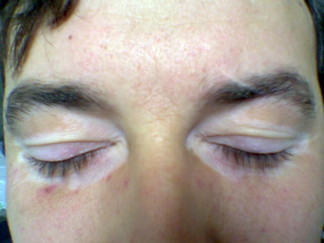
© Maria Sieglinda von Nudeldorf, CC BY-SA 4.0, via Wikimedia Commons
The patches of vitiligo tend to be clearly circumscribed areas of whiteness. They are especially obvious if the surrounding skin is dark. As they will not tan in sunlight they become more apparent after exposure. Furthermore, because of the loss of melanin they are also more susceptible to sunburn.
Lesions are flat and non-scaly. They are often bilateral and symmetrical.
At first there are a few clearly defined lesions that may be slightly darker around the perimeter. They increase in size and number, becoming confluent and it may be difficult, if they are extensive, to decide if it is a dark-skinned person with pale patches or a fair-skinned person with pigmented patches.
Any part of the body can be affected. Most often it is found on the fingers and wrists, neck, nipples, navel and genitalia, and the skin around the eyes and mouth. It may also be found in body folds such as the groin and axillae, and in sites of skin injury. Where vitiligo occurs in sites of injury or friction, it is known as the Köbner phenomenon.
Hair may be white or grey. It is usually in patches on the scalp but can be generalised. Other body hair, including eyebrows and eyelashes, pubic and axillary hair, may also be affected. This is called leukotrichia.
The retina may also be affected.
Variants include trichrome vitiligo in which there is an area of partial depigmentation as well as the depigmented and normal skin, so that there are three colours. There may be marginal inflammatory vitiligo in which a raised red periphery occurs either at onset or up to a year later. Blue vitiligo may occur with post-inflammatory hyperpigmentation that proceeds to vitiligo.
Investigations
The diagnosis is generally made clinically.
Check for evidence of associated disease such as diabetes, pernicious anaemia, thyroid disease and Addison's disease. Consider blood tests for thyroid function and thyroid autoantibodies.
A Wood's light can be helpful to exclude superficial fungal infections that fluoresce in the ultraviolet light. Not all fungal infections fluoresce and the colour with which they fluoresce also varies. If a Wood's light is shone on areas of depigmentation, the exact margin is more readily seen on fair skin and the lesions appear a bright blue-white.
Assess the impact on quality of life.
Differential diagnosis6
Tinea versicolor. Lesions are dry and slightly scaly when scratched.
Piebaldism. A rare genetic condition. There is often a white forelock, which may be present at birth.
Idiopathic guttate hypomelanosis. Numerous small white macules (measuring 1-5 mm) are distributed symmetrically on the trunk, arms and legs. Lesions have well-defined borders and normal skin markings. It has a different appearance under a Wood's light.
Use of potent topical steroids or other types of scarring.
Post-inflammatory hypopigmentation, following inflammatory skin conditions such as eczema.
Pityriasis alba. This may be a type of eczema or an inflammatory reaction following mild eczema.
Leprosy, especially the tuberculoid variety.
Lichen sclerosus and atrophicus. Itchy white patches on the perineum most frequently.
Melasma (chloasma). Hyperpigmentation of the face in women who are pregnant or on the combined oral contraceptive pill.
Albinism. Generalised lack of pigment, including the eyes, present from birth.
Morphoea. Localised thickening of the dermis due to excess collagen.
Vitiligo treatment6 9
The variety of treatments available suggests that none is totally satisfactory. The response is highly variable between patients. A Cochrane review in 2015 concluded it was not possible to establish which was the best treatment for vitiligo and that further studies are needed.5
General measures7
Protect against sun exposure, as white patches can only burn and cannot tan. Advise about wearing protective clothing, avoiding the sun at peak sun times, avoiding the use of sunbeds, and use of high-factor sunscreen. High-factor sunscreens can be prescribed on the NHS if endorsed ACBS. On a more cosmetic note, tanning of normal skin makes the patches of vitiligo more apparent.
Minimise skin injury, as there is an increased likelihood of new white patches in areas of injured skin.
Monitor for other autoimmune conditions in adults with NSV. Look for symptoms of diabetes, pernicious anaemia, Addison's disease and thyroid disease. Advise people with vitiligo to report symptoms of these conditions. Blood tests for thyroid function and thyroid autoantibodies should be done at diagnosis and then annually thereafter.
Assess impact on quality of life and presence of psychosocial problems.
Specific treatment is known to be more effective if started early, when the affected area is small, and in childhood.
If vitamin D levels are reduced or deficient, particularly in view of the advice for reduced sun exposure, advise supplementary vitamin D3 and increasing intake of foods high in vitamin D. See also the article on Vitamin D Deficiency.
Camouflage options
Refer to Changing Faces.10 This provides education to the patient and advice for GPs about the appropriate cream to prescribe.
Highly pigmented cover creams and powders are available in a range of shades and colours that can be colour matched to the person's skin tone. They are waterproof, and easy to apply to the face and anywhere on the body after training. They may remain on the face for 12–18 hours, and on the body for up to 4 days. The camouflage products Covermark® classic foundation and finishing powder, Dermacolor® cream and fixing powder, Keromask® masking cream and finishing powder, and Veil® cover cream and finishing powder are available in different shades and can be prescribed on the NHS (classified as 'borderline substances' and the prescription must be endorsed 'ACBS').
Alternative options include self-tanning products that provide lasting colour for up to several days. Cosmetic micropigmentation and tattoo is a more permanent option, for example for depigmented lips or nipples, however, this should be considered with caution due to the unpredictable course of vitiligo.
Topical corticosteroids
These have an anti-inflammatory and immunomodulating effect.
They can be used in children and adults with limited (less than 10% of the body area) non-segmental vitiligo for a maximum of two months. Topical corticosteroids should not be applied to the face and should not be used in pregnant women.
Use a potent topical corticosteroid such as mometasone or betamethasone valerate 0.1%, applied once daily. Discontinue after one month if response is good.
Topical calcineurin inhibitors
Tacrolimus and pimecrolimus creams.
These can be used in adults and children.
They are used for areas of vitiligo on the head and neck.
Initial use is for six months but they can be used for longer if effective. They have a better safety profile than topical corticosteroids.
Phototherapies
Narrow-band ultraviolet B (NB-UVB) phototherapy:
Effective in widespread NSV.
Effective in combination with topical calcineurin inhibitors.
Total body treatment is recommended for lesions more than 15-20% of body area.
Used in children and adults who have widespread vitiligo, or localised vitiligo which cannot be managed with topical treatments or which is having a significant impact on quality of life.
PUVA (psoralen plus UVA radiation):
Oral or topical psoralen followed by exposure to long-wave UV light for a few minutes.
It is more effective on the face and trunk.
Treatment is twice-weekly for up to two years.
Oral corticosteroids and other immunosuppressants
Some studies show benefit in fast-spreading vitiligo to arrest progress.
On the whole, side-effects and risks outweigh benefit and they are not commonly used.
Surgical treatments
The top layer of vitiligo skin is removed by shaving, dermabrasion or laser and replaced with pigmented skin.
For areas where there have been no new lesions or spreading of lesions in the previous 12 months, and no Köbner phenomenon (skin lesions on lines of trauma).
Surgical treatment options include:
Non-cultured melanocyte-keratinocyte cell transplantation.
Punch grafts.
Blister grafts.
Split skin grafts.
Depigmentation therapy
This is an option for dark-skinned people, who have large areas of skin affected.
The normal skin is treated to depigment it so that all skin is the same colour.
A monobenzone ethyl ester cream is applied.
Treatment takes 1-4 months to work.
It is usually permanent.
Treatment in Primary Care6
General measures as above.
Consider and discuss the option of no treatment where appropriate.
Offer prescriptions for high-factor sun cream and camouflage creams where appropriate.
Referral to the Changing Faces camouflage service.10
Give information on the Vitiligo Society for support.11
Consider treatment with a topical corticosteroid if:
NSV is localised or limited.
The facial area is not involved.
The patient is not pregnant.
Risks are accepted.
No treatment is not an option.
The patient is an adult.
Indications for referral
Consider arranging referral to a dermatologist if:
The condition is progressing rapidly.
The diagnosis is uncertain.
The person has segmental vitiligo.
The face is affected.
A child.
Pregnant.
Large areas of the body are affected (more than 10% of the body surface area).
The person is particularly distressed by the condition.
There are contraindications or adverse effects from topical corticosteroid treatment.
Initial treatment in primary care has been unsuccessful.
Consider arranging referral to a local skin camouflage service (which may be available through the local dermatology service). Advise can self-refer to the charity Changing Faces, which provides education from skin camouflage practitioners on the use and application of cosmetic camouflage creams and powders (see link in References below).
Exclusive updates for healthcare professionals
Stay informed with the latest clinical updates, professional insights, and evidence-based guidance. The Patient Pro newsletter curates essential content for healthcare professionals—delivered straight to your inbox.
By subscribing you accept our Privacy Policy. You can unsubscribe at any time. We never sell your data.
Further reading and references
- Vitiligo; Primary Care Dermatology Society (PCDS)
- Bergqvist C, Ezzedine K; Vitiligo: A Review. Dermatology. 2020;236(6):571-592. doi: 10.1159/000506103. Epub 2020 Mar 10.
- Sandru F, Carsote M, Albu SE, et al; Vitiligo and chronic autoimmune thyroiditis. J Med Life. 2021 Mar-Apr;14(2):127-130. doi: 10.25122/jml-2019-0134.
- Laddha NC, Dwivedi M, Mansuri MS, et al; Vitiligo: interplay between oxidative stress and immune system. Exp Dermatol. 2013 Apr;22(4):245-50. doi: 10.1111/exd.12103. Epub 2013 Feb 21.
- Glassman SJ; Vitiligo, reactive oxygen species and T-cells. Clin Sci (Lond). 2011 Feb;120(3):99-120. doi: 10.1042/CS20090603.
- Whitton ME, Pinart M, Batchelor J, et al; Interventions for vitiligo. Cochrane Database Syst Rev. 2015 Feb 24;(2):CD003263. doi: 10.1002/14651858.CD003263.pub5.
- Vitiligo; NICE CKS, January 2022 (UK access only)
- Vitiligo; DermNet NZ
- Ezzedine K, Eleftheriadou V, Whitton M, et al; Vitiligo. Lancet. 2015 Jul 4;386(9988):74-84. doi: 10.1016/S0140-6736(14)60763-7. Epub 2015 Jan 15.
- Nahhas AF, Braunberger TL, Hamzavi IH; Update on the Management of Vitiligo. Skin Therapy Lett. 2019 May;24(3):1-6.
- Changing Faces
- Vitiligo Society UK
About the authorView full bio

Dr Colin Tidy, MRCGP
General Practitioner, Medical Author
MBBS, MRCGP, MRCP (Paediatrics), DCH
Dr Colin Tidy is an NHS Doctor, based in Oxfordshire.
About the reviewerView full bio

Dr Doug McKechnie, MRCGP
Medical Writer
MA, MBBS, MSc, DRCOG, MRCP(UK), MRCGP(2021), FHEA
Dr Doug McKechnie is an NHS GP working in London. He works full-time clinically and is also the Deputy Lead for the Clinical and Professional Practice module at University College London Medical School.
Article history
The information on this page is written and peer reviewed by qualified clinicians.
Article also available in English, German, Spanish, French, Italian, Portuguese, Hindi, Hebrew, Arabic, and Swedish.
Next review due: 15 Nov 2027
15 Dec 2022 | Latest version

Ask, share, connect.
Browse discussions, ask questions, and share experiences across hundreds of health topics.

Feeling unwell?
Assess your symptoms online for free
More in dermatology
- Allergic phenomena
- Blue naevus
- Cicatricial pemphigoid
- Eczema on hands and feet
- Generalised pustular psoriasis
- Haemangiomata of skin
- Keloid scars
- Keratosis pilaris
- Lentigo
- Orbital and preseptal cellulitis
- Pellagra
- Pressure ulcers
- PUVA
- Rubella
- Sarcoidosis
- Scabies
- Smallpox
- Squamous cell carcinoma of skin
- Staphylococcal scalded skin syndrome
- Stevens-Johnson syndrome