Vitreous haemorrhage
Peer reviewed by Dr Doug McKechnie, MRCGPLast updated by Dr Colin Tidy, MRCGPLast updated 29 Jun 2024
Meets Patient’s editorial guidelines
- DownloadDownload
- Share
- Language
- Discussion
- Audio Version
- Add to preferred sources on Google
In this series:Visual problemsMacular degenerationEye floaters, flashes and haloesRetinal vein occlusionGiant Cell ArteritisSquint in children
Vitreous haemorrhage is bleeding into the jelly-like filling of the back part of your eye. This substance is the vitreous humour. It helps the eye keep its shape and is normally clear, allowing light from outside the eye to pass through it to reach the retina.
Vitreous haemorrhage varies in degree from mild, with 'floaters' and haziness in the vision, to complete loss of vision. It is painless and it comes on quite quickly. Usually only one eye is affected. Whilst it is very alarming, once the bleeding has been treated, many cases resolve and vision is restored to where it was before.
At a glance
Vitreous haemorrhage is when blood leaks into the jelly-like substance that fills your eye.
This can cause symptoms from floaters and hazy vision to complete loss of sight.
It is often caused by diabetic eye disease, retinal tears, or eye trauma.
Diagnosis involves an eye examination and sometimes an ultrasound scan.
Treatment depends on the cause and may involve laser treatment or surgery.
If you have sudden vision loss, see a GP or optician, or go to A&E immediately.
What is vitreous haemorrhage?
Vitreous haemorrhage occurs when blood leaks into the vitreous humour inside the eye. The leaked blood most commonly comes from blood vessels at the back of the eye (retinal blood vessels). This is more likely to happen if the blood vessels have been damaged (eg, by trauma) or are particularly fragile (because of eye disease related to diabetes).
In order for us to see clearly, the vitreous humour needs to be clear. If the vitreous humour is clouded or filled with blood, vision will be impaired. This impairment varies from a few 'floaters' and cloudiness of the vision through to the vision going completely dark (sometimes with a reddish tinge).
Vitreous haemorrhage can therefore cause anything from floaters, hazy or dulled vision (reduced visual acuity) to complete loss of vision.
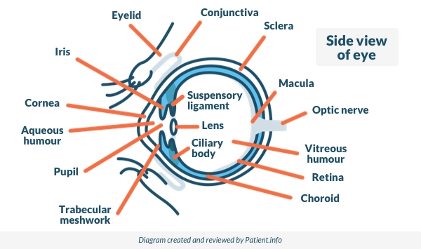
What is the vitreous humour?
The vitreous humour is a clear jelly-like substance that makes up about 80% of the volume of the globe of the eye. It supports the shape of the globe of the eye whilst letting light through.
It is made up mainly of water but with some collagen and hyaluronic acid. The outside is made of fine fibres which are attached to the retina at the back and to the back of the lens at the front.
Side View of the Eye
What are the causes of vitreous haemorrhage?
The most common causes, accounting for about 90% of all cases of vitreous haemorrhage, are:
Bleeding from abnormal new blood vessels forming in advanced diabetic eye disease.
Bleeding from tears in the retina caused by vitreous detachment (see below).
Trauma to the eye (the most common cause in younger people).
Bleeding inside the eye can come from:
Abnormal blood vessels which grow because the back of the eye is short of oxygen. These are fragile and bleed easily. Conditions in which this can occur include:
Diabetic eye disease (the most common cause).
Retinopathy with sickle cell disease.
Damage to the back of the eye in very premature babies who have been on oxygen in special care baby units.
Normal blood vessels which are damaged. They may be damaged by:
Posterior vitreous detachment, often because it causes a retinal tear (see below).
Retinal macroaneurysms - swollen blood vessels on the retina, usually related to high blood pressure, atherosclerosis and smoking.
Blunt trauma - suddenly compressing the eye - for example, if hit by a squash ball.
Penetrating trauma - this will cause bleeding throughout the eye. Penetrating trauma can occur from high-velocity injuries such as grinding and hammering. They do not always cause severe eye pain.
Subarachnoid haemorrhage, which can increase the pressure in the veins in the retina, causing them to bleed.
Surgery to the eye, particularly if it involves the inside of the eye.
Blood from behind the retina tracking through into the eye. This is the least common cause of vitreous haemorrhage. It may result from:
Tumours of the back of the eye (eye tumours are rare. The most common type is ocular melanoma).
Fragile new blood vessels behind the retina.
Posterior vitreous detachment is usually experienced between the ages of 60 and 80 - it happens to most of us between those ages. It occurs when the vitreous pulls away from the retina at the back. This can happen quite suddenly as the vitreous tends to shrink with age.
Most commonly there are no symptoms. Sometimes posterior vitreous detachment causes bleeding from the retina as it pulls away. Sometimes the retina is torn as the vitreous pulls away from it - in this case bleeding is more likely.
Most people experience posterior vitreous detachment in their 60s or later, and most do not have significant vitreous haemorrhage when it happens. However, episodes of noticeable floaters are very common and are probably caused by tiny bleeds.
Vitreous haemorrhage symptoms
The symptoms of smaller bleeds (most bleeds are smaller bleeds) include:
Floaters in vision.
Cobweb haze.
Haze and shadows in the eye.
Red tint to the vision.
Symptoms most commonly affect one eye only, although both eyes can be affected.
Some people find that their vision is worse in the morning, because blood settles to the back of the eye overnight whilst lying down.
More severe bleeds cause:
Haziness of vision.
Blind spots or dark streaks.
The most severe bleeds cause visual loss, which can be complete, leaving the vision hazily red or black. For most people this is extremely alarming, particularly as it tends to come on very quickly with no clear explanation.
How common is vitreous haemorrhage?
Vitreous haemorrhage affects about 7 per 100,000 people each year. This makes it one of the most common causes of sudden deterioration in vision. It most often affects only one eye.
Who is likely to experience vitreous haemorrhage?
The most common cause of vitreous haemorrhage is severe diabetic eye disease, which is mainly seen in older adults. The other common causes of vitreous haemorrhage also tend to occur in adults aged 60 and above, except for eye trauma, which can occur at any age.
How is vitreous haemorrhage diagnosed?
An examination using a slit lamp is performed by opticians and eye specialists (ophthalmologists) to look in detail at the inside of the eye. The slit lamp will allow the examiner to see if there is blood in the vitreous.
Finding the source of the bleeding may be possible with the slit lamp, although if there is a lot of blood in the vitreous humour this prevents a clear view and it may therefore be difficult to know what has happened. In this case an ultrasound scan of your eye can help. Ultrasound can detect many causes of vitreous haemorrhage, including posterior vitreous detachment, retinal tears and detachments, tumours and foreign objects.
Sometimes an angiogram is needed. This test shows up the blood vessels in the back of the eye. This can be helpful if looking for abnormal blood vessels such as in diabetes.
Computerised tomography (CT) scanning of the eyes is useful if there is a suspicion of a penetrating injury.
What is the treatment for vitreous haemorrhage?
The treatment of vitreous haemorrhage varies with the cause. Aims of treatment are to:
Find the source of the bleeding.
Stop the bleeding.
Repair any damage to the retina before it results in permanent loss of vision.
Restore normal vision.
Once the source of the bleeding has been identified, treatment will depend on the cause. If there is not too much blood in the vitreous and the source of bleeding can be seen then it is treated. This means laser treatment to bleeding vessels and any other abnormal vessels, and repair to any tears in the retina. After this it is a matter of waiting for the blood to slowly clear as the red blood cells break down. This can take several weeks.
Strenuous activity should be avoided for several days at least, as this might dislodge clots and trigger new bleeding. It is also advised to sleep with the head of the bed elevated, as this allows the blood in the vitreous to settle into the bottom of the eye, out of the line of vision.
If the blood in the vitreous obscures the view and prevents treatment of the bleeding then the entire vitreous may be removed first. This procedure is called a vitrectomy. Doctors will perform a vitrectomy if they can't see the back of the eye, or if the view isn't good enough to treat the bleeding there safely.
Specific treatments
Laser photocoagulation
This is the usual treatment for fragile abnormal vessels. Treating them both stops the bleeding and prevents later bleeding. Laser photocoagulation is also used in repairing damage to the retina, including retinal detachments.
Anti-VEGF injections
Anti-VEGF injections (a type of intravitreal injection) aim to shrink abnormal new vessels which have formed in the eye. They are sometimes used in patients with diabetes, in addition to other treatments like laser photocoagulation and vitrectomy, in order to reduce bleeding.
Cryotherapy
This is also used as a treatment for retinal tears and retinal detachments.
Vitrectomy
Vitrectomy is removal of the vitreous humour completely, together with the membrane that surrounds it. This is done when there is so much blood in the vitreous that it is impossible to diagnose and treat the cause. Vitrectomy is also sometimes performed if the blood in the vitreous is clearing very slowly and vision remains impaired.
Waiting
Waiting is commonly the chosen option once the bleeding has stopped. Most cases of vitreous haemorrhage do not require vitrectomy. The blood clears slowly from the vitreous, allowing light to pass through it again. If the underlying vision is undamaged then normal vision will be restored.
General measures
Anyone with a suspected vitreous haemorrhage will usually be seen by an eye specialist on the same day. This is because sudden loss of vision is considered an eye emergency. The aim is to ensure accurate diagnosis and to avoid permanent loss of vision which could occur if there is a retinal detachment behind the bleeding.
Anyone who has a sudden loss of vision or any other unexplained disturbance in vision in one or both eyes should:
See a GP or optician, or attend an accident and emergency department, as soon as possible.
Try to remain calm - most vitreous haemorrhages respond well to treatment and there is a very good chance that vision will return to where it was.
Rest for a few hours a day sitting upwards and elevate the head at night on pillows.
Ask a GP to check blood pressure - raised blood pressure increases the chance of further bleeding.
Avoid any heavy lifting, which can increase the chance of further bleeding.
What is the outlook for vitreous haemorrhage?
The outlook (prognosis) in vitreous haemorrhage depends both on the cause and on the severity.
Vitreous haemorrhage resulting from posterior vitreous detachment usually has a good prognosis, with restoration of vision, particularly if the eye is otherwise normal.
Where severe diabetic eye disease or macular degeneration has resulted in abnormal blood vessels, the outlook for the vision is much less good. The outlook in penetrating eye injury is often poor.
How is vitreous haemorrhage prevented?
Prevention of vitreous haemorrhage involves preventing the underlying causes. This includes careful and regular management of diabetic eye disease (which tends to be worse in less well-controlled diabetes) and high blood pressure, and giving up smoking.
The eye should always be protected from trauma during high-risk activities such as filing, grinding and hammering, using firearms and playing sports with high-speed balls, such as squash.
Dr Mary Lowth is an author or the original author of this leaflet.
Patient picks for Eye conditions

Eye health
Retinal vein occlusion
Retinal vein occlusion occurs when one of the tiny veins in the retina becomes blocked by a blood clot.
by Dr Toni Hazell, FRCGP
%20/og.png)
Eye health
Amblyopia (lazy eye)
Amblyopia (often called a lazy eye) means that vision in one eye does not develop fully during early childhood. Amblyopia is usually a correctable problem if it is treated early. Late treatment can mean that the sight problem remains permanent. A squint (strabismus) is one of the most common causes of amblyopia. Treatment of amblyopia involves making the lazy eye work harder to see. This is usually done by blocking the vision in the good eye with a patch or by making the eyesight in the good eye blurry by using atropine eye drops.
by Dr Philippa Vincent, MRCGP
Frequently asked questions
If I have a vitreous haemorrhage, will I definitely lose my vision permanently?
No, permanent vision loss is not a certainty. The outcome largely depends on the underlying cause and how severe the bleed is. Cases caused by posterior vitreous detachment typically have a good prognosis, often leading to full vision restoration if the eye is otherwise healthy. Prompt diagnosis and treatment are important to prevent permanent vision loss, especially if there's an underlying issue like retinal detachment.
Why is my vision worse in the morning if I have a vitreous haemorrhage?
If you have a vitreous haemorrhage, your vision might seem worse in the morning because when you lie down overnight, the blood settles to the back of your eye. This can obscure your vision more significantly in the line of sight when you wake up.
What happens if the bleeding in my eye clears up on its own without needing surgery?
If the bleeding in your eye clears up on its own, it means the blood in the vitreous is slowly reabsorbing. If the underlying cause of the bleeding is identified and treated (for example, with laser if vessels are visible), and the retina was not damaged, then your normal vision should be restored over time as the blood clears completely.
Is vitreous haemorrhage a common condition?
Vitreous haemorrhage affects about 7 out of every 100,000 people each year, making it one of the more common reasons for sudden vision deterioration. It usually impacts only one eye.
Can I prevent a vitreous haemorrhage from happening again after I've had one?
Preventing a recurrence of vitreous haemorrhage involves managing the underlying conditions that caused it. This means carefully and regularly controlling conditions like diabetic eye disease and high blood pressure, as well as quitting smoking. For high-risk activities such as grinding, hammering, or playing sports with fast-moving balls, it's advised to always protect your eyes from trauma.
Does being older make me more likely to get a vitreous haemorrhage?
Yes, older age can increase your likelihood of experiencing a vitreous haemorrhage. The most common cause, severe diabetic eye disease, mainly affects older adults. Other common causes, apart from eye trauma, also tend to occur in people aged 60 and above.
What is the typical recovery time after a vitrectomy for vitreous haemorrhage?
The article states that once the bleeding has stopped, it can take several weeks for blood to slowly clear from the eye. However, it does not specify a separate recovery time directly related to the vitrectomy procedure itself, beyond the clearing of blood.
Further reading and references
- Shaikh N, Srishti R, Khanum A, et al; Vitreous hemorrhage - Causes, diagnosis, and management. Indian J Ophthalmol. 2023 Jan;71(1):28-38. doi: 10.4103/ijo.IJO_928_22.
- Mathen,T, Leng, T, Shah VA; Terson Syndrome, EyeWiki - American Academy of Ophthalmology
About the authorView full bio

Dr Colin Tidy, MRCGP
General Practitioner, Medical Author
MBBS, MRCGP, MRCP (Paediatrics), DCH
Dr Colin Tidy is an NHS Doctor, based in Oxfordshire.
About the reviewerView full bio

Dr Doug McKechnie, MRCGP
Medical Writer
MA, MBBS, MSc, DRCOG, MRCP(UK), MRCGP(2021), FHEA
Dr Doug McKechnie is an NHS GP working in London. He works full-time clinically and is also the Deputy Lead for the Clinical and Professional Practice module at University College London Medical School.
Article history
The information on this page is written and peer reviewed by qualified clinicians.
Article also available in English, German, Spanish, French, Italian, Portuguese, Hindi, Hebrew, Arabic, and Swedish.
Next review due: 28 Jun 2027
29 Jun 2024 | Latest version

Ask, share, connect.
Browse discussions, ask questions, and share experiences across hundreds of health topics.

Feeling unwell?
Assess your symptoms online for free
Sign up to the Patient newsletter
Your weekly dose of clear, trustworthy health advice - written to help you feel informed, confident and in control.
By subscribing you accept our Privacy Policy. You can unsubscribe at any time. We never sell your data.
More in eye health
- Amblyopia (lazy eye)
- Astigmatism
- Colour vision deficiency
- Contact lenses
- Dry eyes
- Entropion
- Eye floaters, flashes and haloes
- Eye problems
- Giant Cell Arteritis
- Hypermetropia
- Idiopathic intracranial hypertension
- Myopia
- Retinal artery occlusion
- Retinal vein occlusion
- Sjögren's syndrome
- Squint in children
- Stye
- Subconjunctival haemorrhage
- Swollen eyelid